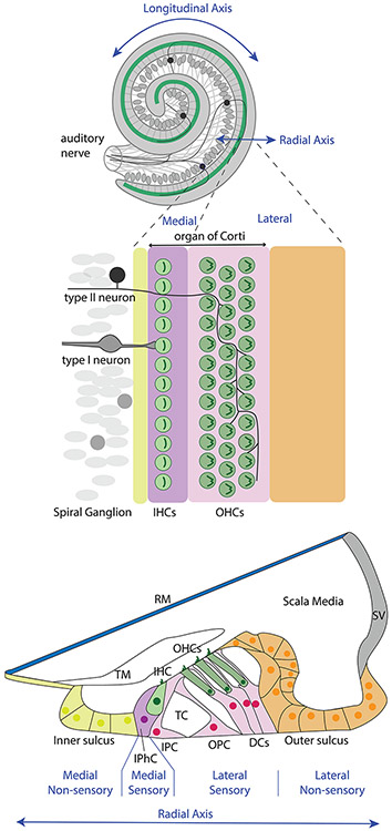
Figure 1. Cochlear anatomy and connectivity.
Inner hair cells reside on the “medial” side of the tunnel of Corti. This is the direction closer to the center (or middle) of the cochlear coil, where axons entering and leaving the cochlea through the auditory nerve are bundled together. The IHC bodies are arranged in a row above the nuclei of their associated supporting cells, called inner phalangeal cells. They are contacted by the peripheral processes of neurons residing in the nearby spiral ganglion. The overwhelming majority of spiral ganglion neurons, the type I neurons, each make a synapse with a just one IHC. Each IHC makes synapses with 20-30 spiral ganglion neurons (not shown). The stereocilia bundles of the IHCs are displaced by fluid movement within the scala media. Sensory input to the brain thus begins at the hair cell, travels across the chemical synapse to depolarize the neuron’s peripheral process, which generates an action potential that travels along the neuron’s central process into the cochlear nucleus of the brain. Once this axon enters the brain, it branches extensively to make chemical synapses with (i.e., to innervate) a variety of cell types and subdivisions of the cochlear nucleus (not shown). Just lateral to the IHCs are the inner and outer pillar cells that create the tunnel of Corti. The pillar cells are placed within the “lateral” side of the organ of Corti, which also included three rows of OHCs and the Deiter’s cells that support them. The OHCs are stimulated by the mechanical displacement of the stereocilia that are in contact with the tectorial membrane upon movement of the basolateral membrane. OHCs receive innervation from the small population of type II spiral ganglion neurons whose peripheral processes cross the tunnel, turn basally and branch extensively. The medial and lateral sides of the organ of Corti also receive efferent innervation from distinct populations of neurons located in the brainstem (not shown). Overall, the differences in neural circuitry and organ of Corti cellular phenotypes emphasize and impart very different functions to the two halves of the organ of Corti’s radial axis. Abbreviations: DCs, Deiter’s cells, IHC, inner hair cell; IPC, inner pillar cell; IPhC, inner phalangeal cell, OHCs, outer hair cells; OPC, outer pillar cell; RM, Reissner’s membrane; SV, stria vascularis; TC, tunnel of Corti, TM, tectorial membrane.

