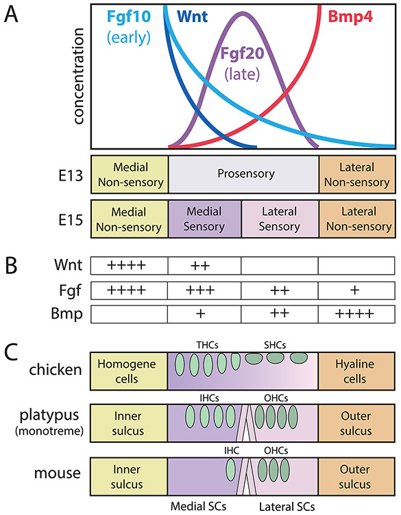
Figure 3. A model for how protein gradients across the floor of the cochlear duct may contribute to its segregation into compartments.
(A) Protein concentrations (y-axis) are presumed to be distributed as gradients across the radial axis from medial (left) to lateral (right). This schematic is representative of the mouse cochlea during the window of embryonic days 11.5-15.5 (see Groves and Fekete, 2012, for a more explicit temporal sequence). These proteins are encoded by mRNA transcripts (not shown) that end rather abruptly at borders of the prosensory domain, thus providing asymmetric sources of proteins that may function as morphogens. (B) Within the prosensory domain, exposure to specific levels (+, ++, +++) of the major signaling factors could serve to subdivide it into medial and lateral compartments. (C) Later, within each compartment, each cell has acquired positional information that instructs its eventual fate across the radial axis of the organ of Corti. Domains are shown as different colors; oval-shaped hair cells shown in green; pillar cells shown as vertical rods. In different species, the radial pattern that arises is homologous but not identical to the mammal, when considering cell types or their relative numbers. This is probably due to differences in the shape of the morphogen gradients, along with evolutionary modifications of the precise developmental programs that are activated in response to morphogen exposure. Abbreviations: IHCs, inner hair cells; OHCs, outer hair cells; SCs, supporting cells; SHCs, short hair cells; THCs, tall hair cells.

