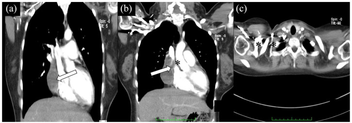
Figure 2.
CT scan shows distinct hematoma (arrows) without any sign of compression of cardiac structures (a). Three months after surgery, expanding pericardial hematoma compressing the SVC, extending anterosuperiorly to the level of the aortic arch (b). Collateral vessels were also visualized (c).
Note: Asterisk showing vein compression.

