
Figure 7. Subtelomeric DNA replicates early in human male meiosis.
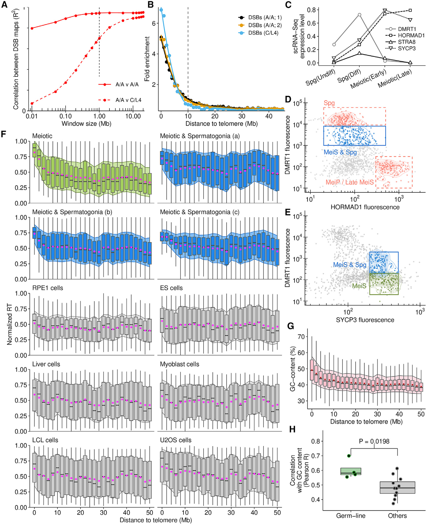
(A) DSB density correlates at megabase-scale in human males with different PRDM9 genotypes (PRDM9A/A homozygotes [A/A] and PRDM9C/L4 [C/L4] heterozygote).
(B) Points represent the average DMC1-SSDS signal in 1-Mb bins, normalized to interstitial regions (>30 Mb).
(C) Stage-specific expression of meiotic markers (Guo et al., 2018).
(D) Putative meiotic S-phase nuclei gated on DMRT1 and HORMAD1 (blue) contain spermatogonia (manual inspection).
(E) A pure meiotic S-phase population (green) is isolated using DMRT1 and SYCP3.
(F) Subtelomeric DNA replicates consistently early in meiotic (green, blue) but not in somatic cells (gray; Table S3). Boxplots depict the interquartile range of replication timing values in 2-Mb regions across the genome; gray bar, median; magenta dot, mean ± 1 SD (filled shadow).
(G) GC content is elevated in subtelomeric DNA.
(H) RT in the germline (all meiotic samples) correlates better with genomic GC content than RT in other cell types.
See also Data S2.