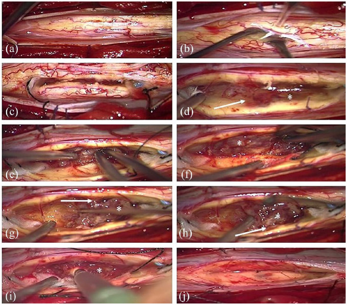
Figure 2.
The spinal cord is exposed after dura opening and bulged due to the intramedullary tumor (a). Myelotomy performed medially (b). The cranial and caudal boundary (see tidal flats) of the tumor is prepared (c). The margins (→) of the grayish tumor (*) are well defined (d). Debulking of the tumor and piecemeal removal using a CUSA with preservation of the surrounding spinal cord tissue (e–i). Spinal cord after complete tumor removal (j).

