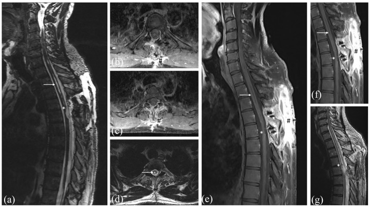
Figure 3.
Early postoperative T2-weighted MRI without contrast (a + d) and T1-weighted MRI with contrast (b + c) 24 h after surgery with completely removed ependymoma. T1-weighted MRI with contrast (e + f) 6 months after surgery showing no tumor recurrence (*) despite the normal contrast enhancement at the dorsal approach (#). T2-weighted MRI without contrast showing the postoperative changes of the spinal cord (*) and the smaller edema of the spinal cord (→) (g).

