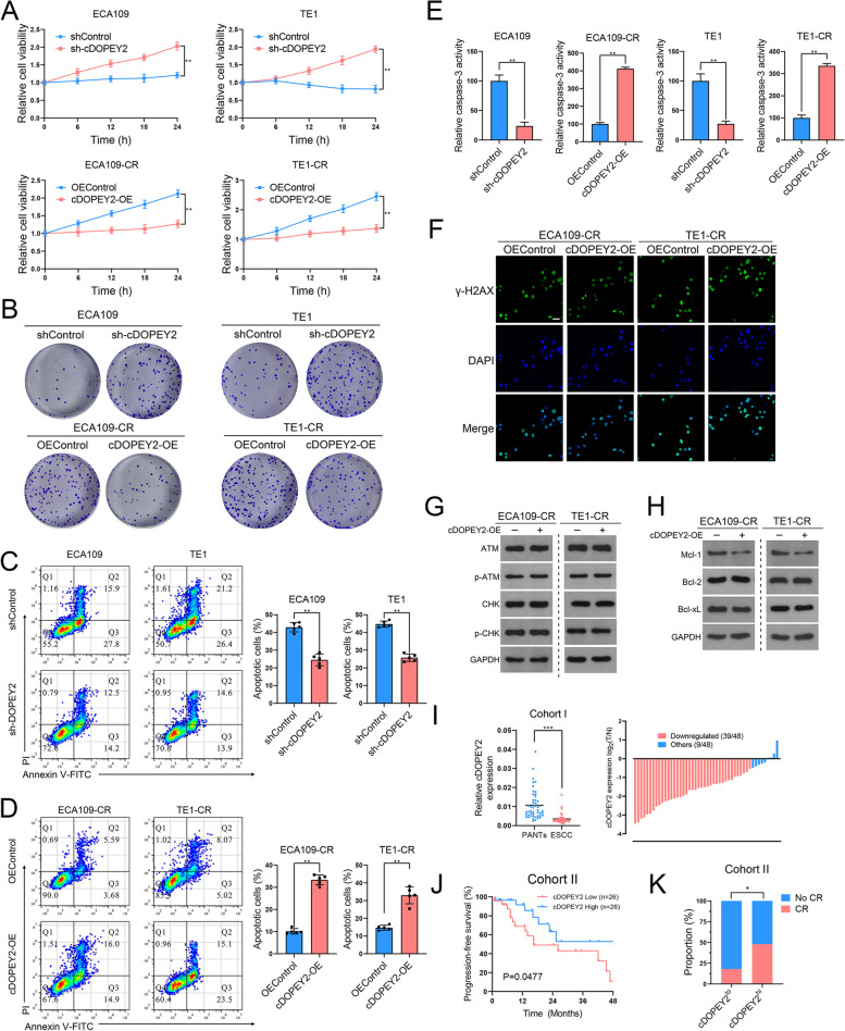
Fig. 2.
cDOPEY2 attenuates cisplatin resistance in ESCC cells. A The viability of the indicated cells with cDOPEY2 overexpression or knockdown after treatment with cisplatin (10 µg/mL) was measured by the CCK-8 method. B The impact of cDOPEY2 on ESCC cells treated with cisplatin was determined by a clonogenic assay. C-D The apoptotic rate of the indicated cells with cDOPEY2 overexpression or knockdown after treatment with cisplatin (10 µg/mL) for 24 h was determined by Annexin V-FITC/PI FACS analysis. E Relative caspase-3 activity in the indicated ESCC cells 24 h after cisplatin treatment at a dose of 10 µg/mL. F Representative γ-H2AX staining images of the indicated ESCC cells 24 h after cisplatin treatment at a dose of 10 µg/mL. Scale bars: 20 μm. G-H Western blot analysis of the indicated proteins in cDOPEY2-overexpressing and cDOPEY2-silenced cells 24 h after cisplatin treatment at a dose of 10 µg/mL. I Relative expression of cDOPEY2 in 48 paired ESCC samples in cohort (I) GAPDH was used as an internal control. J Kaplan-Meier (K-M) plot showing the relationship between cDOPEY2 expression and patient progression-free survival (PFS) in cohort (II) K Bar graph showing that cDOPEY2 expression was positively correlated with the complete response rate (CR) among patients receiving cisplatin-based chemotherapy in Cohort II. Data are presented as the mean ± SD. *P < 0.05, **P < 0.01, ***P < 0.001. P values were determined with the unpaired Student’s t-test (A, C, D and E), paired Student’s t-test (I), log-rank test (J), and Fisher’s exact-test (K)

