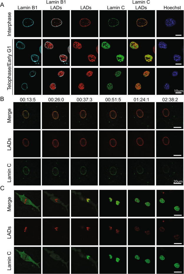
Fig. 4.
Lamin C and LADs do not colocalize but resolve to the lamina at the same time during G1. A Representative images of interphase and early G1 nuclei of anti-lamin B1 (cyan), LAD-tracer (red), lamin C (green), and Hoechst 33342 (blue). B Still images from time lapse movie 3 of LADs (red) and EGFP-LmnC (green) during interphase collected in parallel to panel B. Scale Bar is 20μm. C Still images from time lapse movie 1 of LADs (red), and EGFP-LmnC (green) during mitosis. Scale bar is 20μm. Images were chosen to exemplify certain stages (metaphase, anaphase, telophase, early G1, partially resolved, fully resolved)

