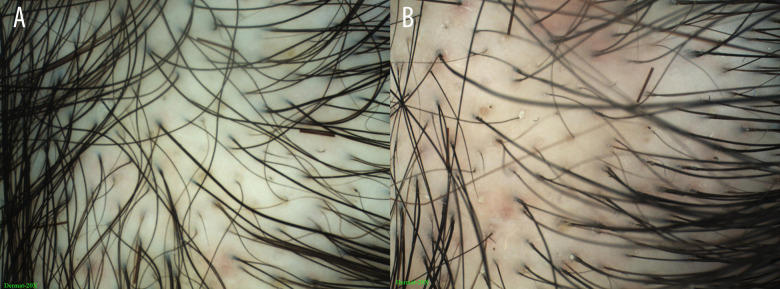
Figure 2.
Dermatoscopy images of a 38-year-old FPHL patient showing shaft variability and yellow spots, created using Dermoscopy-II 2.0 software (Dermat Speedy Recovery T&D Co., Ltd, Beijing, China). (A) Frontal scalp using the Immersion model (20×); (B) Frontal scalp using the Polari-light model (20×).

