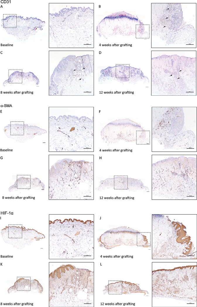
FIGURE 6. Hypoxic signal and revascularization starts from the mouse skin-human skin xenograft interface and grows inwards towards the middle of the graft.

Human CD31 staining of human skin (A) prior to grafting, and at (B) 4 weeks, (C) 8 weeks, and (D) 12 weeks after grafting. Arrows identify CD31 positive vessels. α-SMA staining of human skin (E) prior to grafting, and at (F) 4 weeks, (G) 8 weeks, and (H) 12 weeks after grafting. HIF-1α staining of human skin (I) prior to grafting, and at (J) 4 weeks, (K) 8 weeks, and (L) 12 weeks after grafting. Brown = CD31, α-SMA, HIF-1α antibody, Purple = hematoxylin. Inset boxes (dashed square box) identify the magnified regions. Representative images of three to five animals per time point. Scale bars: 500 μm
