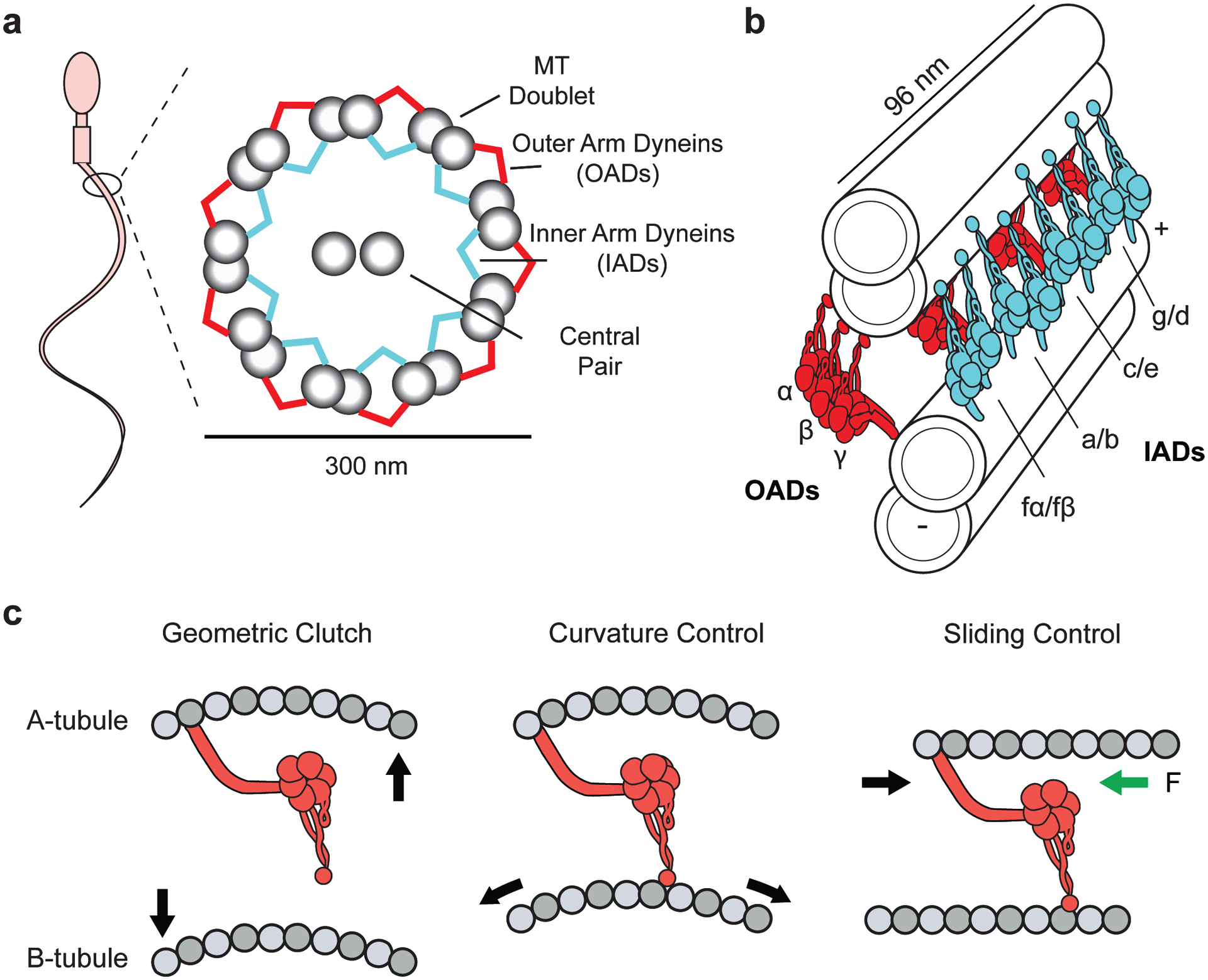
Figure 6.

Structure and regulation of axonemal dyneins. (a) (Left) A schematic representation of a spermatozoon. (Right) Ciliary cross-section displaying the arrangement of dyneins in the inner and outer arm of the axoneme. (b) Organization of OAD (red) and IAD (cyan) motors along adjacent MT doublets. OAD heterotrimers are spaced 24 nm apart with their MTBDs extending toward the adjacent B-tubule. IAD dyad pairs are organized in repeating arrays of (fα–fβ), (a–b), (c–e), (g–d) every 96 nm. (c) Models for dynein regulation during ciliary beating. In the geometric clutch model, ciliary bending changes the distance between adjacent MTs, which prevents the motors from attaching to the MT. In the curvature control model, ciliary bending generates a stretched lattice on the convex side. The OAD MTBD senses the changes in MT curvature. In the sliding control model, nexin linkers generate a resisting force (green arrow) parallel to the axoneme, causing motors to release from the MT at high forces. For clarity, only the transitions from straight to convex bends are displayed. Abbreviations: IAD, inner arm dynein; MT, microtubule; MTBD, MT-binding domain; OAD, outer arm dynein.
