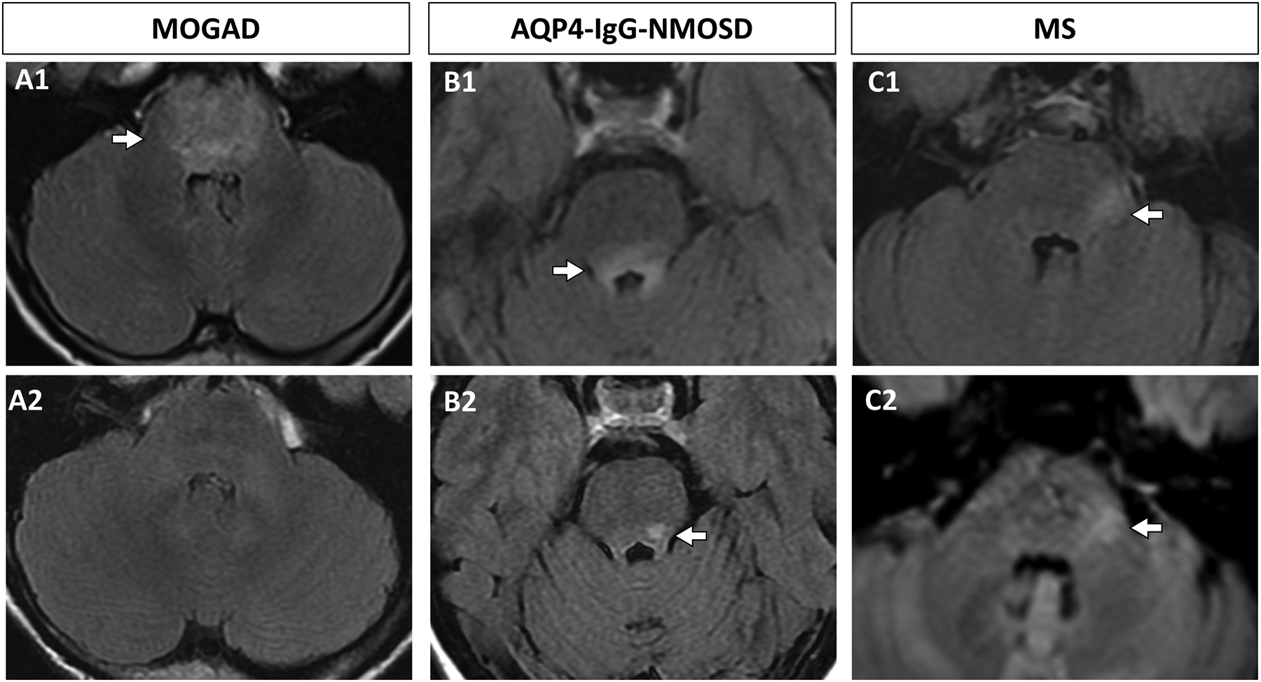
Figure 3. Evolution of brainstem lesions in MOGAD, AQP4-IgG-NMOSD, and MS.

A: A MOGAD pediatric patient had a diffuse T2 FLAIR hyperintense pontine lesion on axial images (A1, arrow) that completely resolved at 6 month follow up (A2). B: An AQP4-IgG-NMOSD seropositive adult patient had a T2 FLAIR hyperintense dorsal pontine lesion on axial images (B1, arrow) that partially resolved at 9 months follow-up (B2, arrow). C: An adult patient with MS had a hyperintense lateral pontine lesion on axial T2 FLAIR images (C1, arrow) that persisted at 11 months follow up (C2, arrow).
Key: AQP4-IgG-NMOSD, aquaporin-4-IgG positive neuromyelitis optica spectrum disorder; FLAIR, fluid-attenuated inversion recovery, MOGAD, myelin oligodendrocyte glycoprotein antibody associated disorder; MS, multiple sclerosis.
