. 2021 Oct 19;10:e66793. doi: 10.7554/eLife.66793

Figure 1. Screening and discovery of a feline skin commensal bacterium that inhibits drug-resistant gram-positive pathogens.

(A) Illustration of the selection and screening strategy of animal-derived staphylococci against the growth of methicillin-resistant S. pseudintermedius (MRSP) ST71 in liquid culture and agar co-culture assays. (B) The panel of 58 feline and canine isolates selected for antimicrobial testing, as well as human-derived S. hominis A9 and S. capitis E12 positive control antimicrobial strains and the non-antimicrobial S. aureus 113 negative control. (C) Inhibition of S. pseudintermedius ST71 growth by OD600, relative to TSB control at 100%, after 18 h incubation in 50%, 25%, or 12.5 % (1:1, 1:4, 1:8 ratio) sterile conditioned supernatant from all staphylococci isolates. Greater than 80 % inhibition of growth was considered antimicrobial (AM+). (D) Images of the agar co-culture assay showing zone of inhibition (black circle surrounding colony) produced by all staphylococci test isolates against S. pseudintermedius ST71, including S. felis C4, N26, V13 (S. f C4, S. f N26, S. f V13), S. pseudintermedius N13 and Q13 (S. p N13 and S. p Q13), and positive control S. hominis A9 (S. h A9, all indicated by arrows). (E) Inhibition of bacterial growth by OD600, normalized to TSB alone at 100%, against select gram-positive and gram-negative pathogens after 18 h incubation (48 h incubation for E. faecium) in the presence of increasing amounts of sterile conditioned supernatant from S. felis C4 overnight growth. Error bars indicate SEM. Representative of three separate experiments.

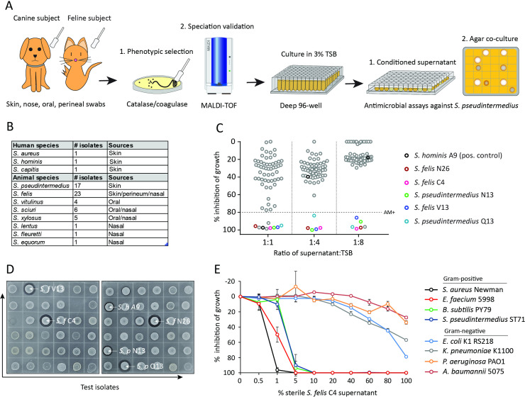
Figure 1—source data 1. Source data for Figure 1C (% growth of S.p ST71 in SN of animal isolates) and Figure 1E (% growth of gram positive and gram negative pathogens in S. felis C4 SN).
Figure 1—source data 2. Source data for Figure 1D (labeled and unlabeled images of the antimicrobial agar assay).

Figure 1.

Figure 1—figure supplement 1. Generation of a partially purified antimicrobial extract from S. felis C4.

Figure 1—figure supplement 1.

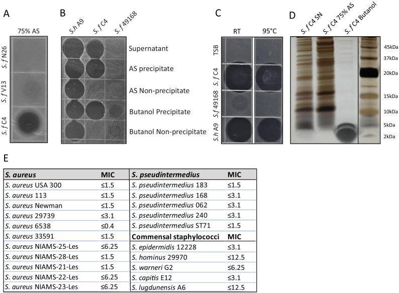
(A) Supernatant of indicated antimicrobial S. felis strains (S. f C4, N26, V13 were incubated with) were incubated with 75 % ammonium sulfate (AS) and the resulting precipitate was inoculated onto agar containing S. pseudintermedius ST71, to determine antimicrobial activity. (B) Supernatant of S. felis C4, positive-control S. hominis A9, or negative-control S. felis ATCC 49168 was extracted in 75 % AS or 25 % n-butanol and activity of the precipitate and non-precipitate fractions were assayed against S. pseudintermedius ST71. (C) Supernatant of S. felis C4 (S. f C4), S. hominis A9 (S. h A9), S. capitis E12 (S. c E12) or TSB alone, were maintained at room temperature (RT) for 1 week or boiled at 95 °C, for 30 min and activity determined by inoculation directly onto agar containing S. pseudintermedius ST71. (D) Total protein silver stain of S. felis C4 supernatant before precipitation or after precipitation in 75 % AS or 25 % butanol. (E) Minimum inhibitory concentration (MIC) table of the S. felis C4 extract against multiple isolates of S. aureus and S. pseudintermedius as well as several common commensal CoNS.
Figure 1—figure supplement 1—source data 1. Source data for Figure 1—figure supplement 1A (labeled and unlabeled images of the antimicrobial agar assay).
Figure 1—figure supplement 1—source data 2. Source data for Figure 1—figure supplement 1B (labeled and unlabeled images of the antimicrobial agar assay).
Figure 1—figure supplement 1—source data 3. Source data for Figure 1—figure supplement 1C (labeled and unlabeled images of the antimicrobial agar assay).
Figure 1—figure supplement 1—source data 4. Source data for Figure 1—figure supplement 1D (labeled and unlabeled images of the silver-stained bacterial protein before and after extraction).
Figure 1—figure supplement 2. S. felis C4 supernatant and extract disrupt S. pseudintermedius biofilm.

Figure 1—figure supplement 2.

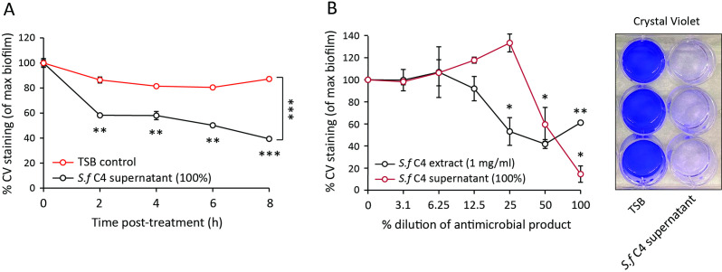
(A) Decrease in crystal violet staining of a 4 h preformed S. pseudintermedius ST71 biofilm during 8 h incubation with 100% S. felis C4 sterile supernatant. (B) Decrease in crystal violet staining of a 4 h preformed S. pseudintermedius ST71 biofilm after 24 h incubation with serially diluted S. felis C4 sterile supernatant (100%) or S. felis C4 extract (1 mg/ml). Right inset is a representative image of crystal violet staining after incubation with 100% S. felis C4 supernatant. (A–B) Error bars indicate SEM. Representative of two separate experiments. A two-tailed, unpaired Student’s t test was performed. p values: *p < 0.05; **p < 0.01; ***p < 0.001.
Figure 1—figure supplement 2—source data 1. Source data for Figure 1—figure supplement 2A,B (raw values for the biofilm inhibition).