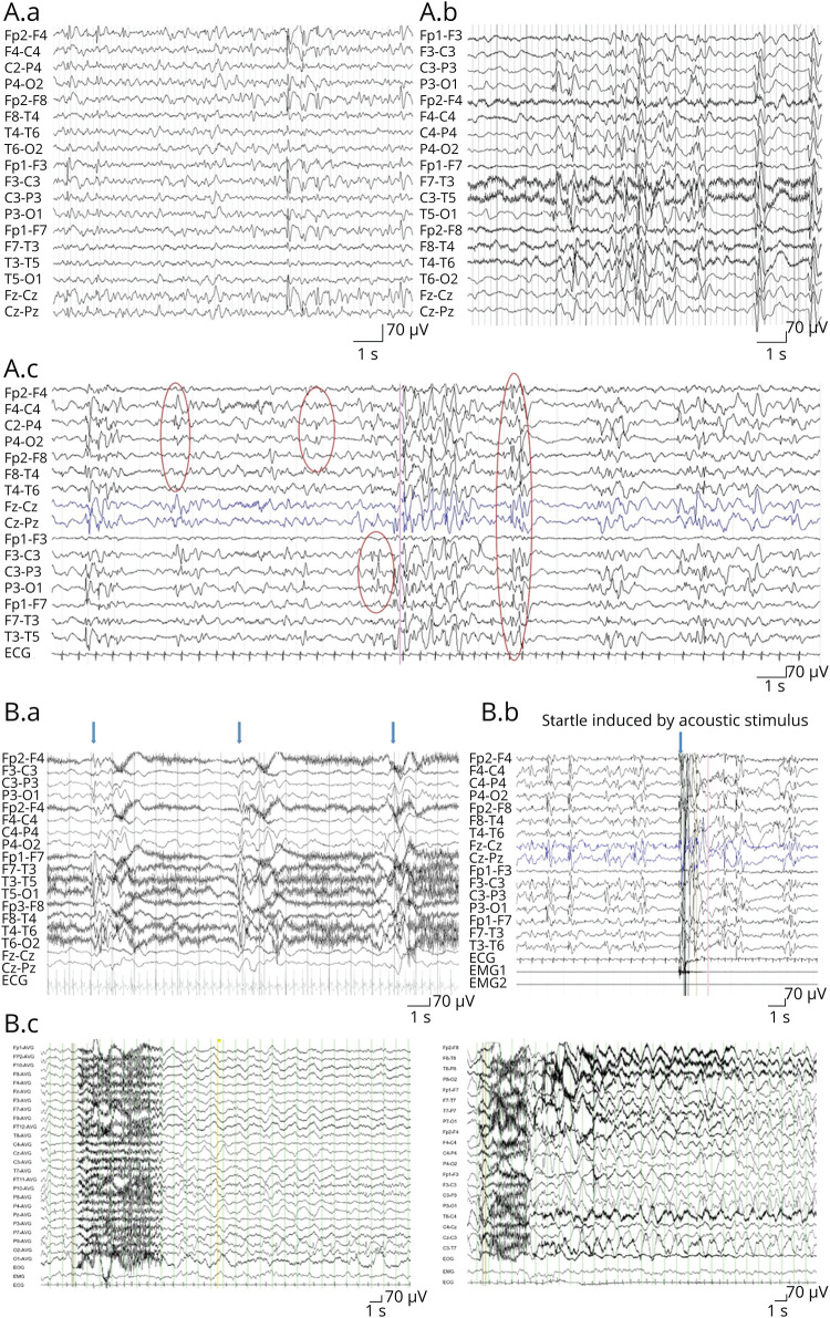
Figure 2. EEG Changes in the PURA Syndrome.
(A) The interictal EEG is characterized by slow background and multifocal spike/sharp and slow waves predominant either in the frontal regions (a, pt. #42) or in the posterior quadrants (b, pt. #30). The epileptiform activity is accentuated during sleep; this image (c, pt. #19) shows very frequent multifocal spike/polyspikes and waves and sharp and slow waves, independently in the right central, right parietal, and left central regions or diffuse. (B) The ictal EEG showed (a) cluster of epileptic spasms (each arrow corresponds to a spasm) (pt. #30), (b) a startle induced by acoustic stimulus (pt. #19), and (c) brief tonic seizures out of sleep with an EEG correlation consisting of diffuse rapid activity (3–6 seconds) followed by diffuse delta activity and trains of spike and slow waves in the posterior regions (pt. #30). PURA = purine-rich element-binding protein A.

