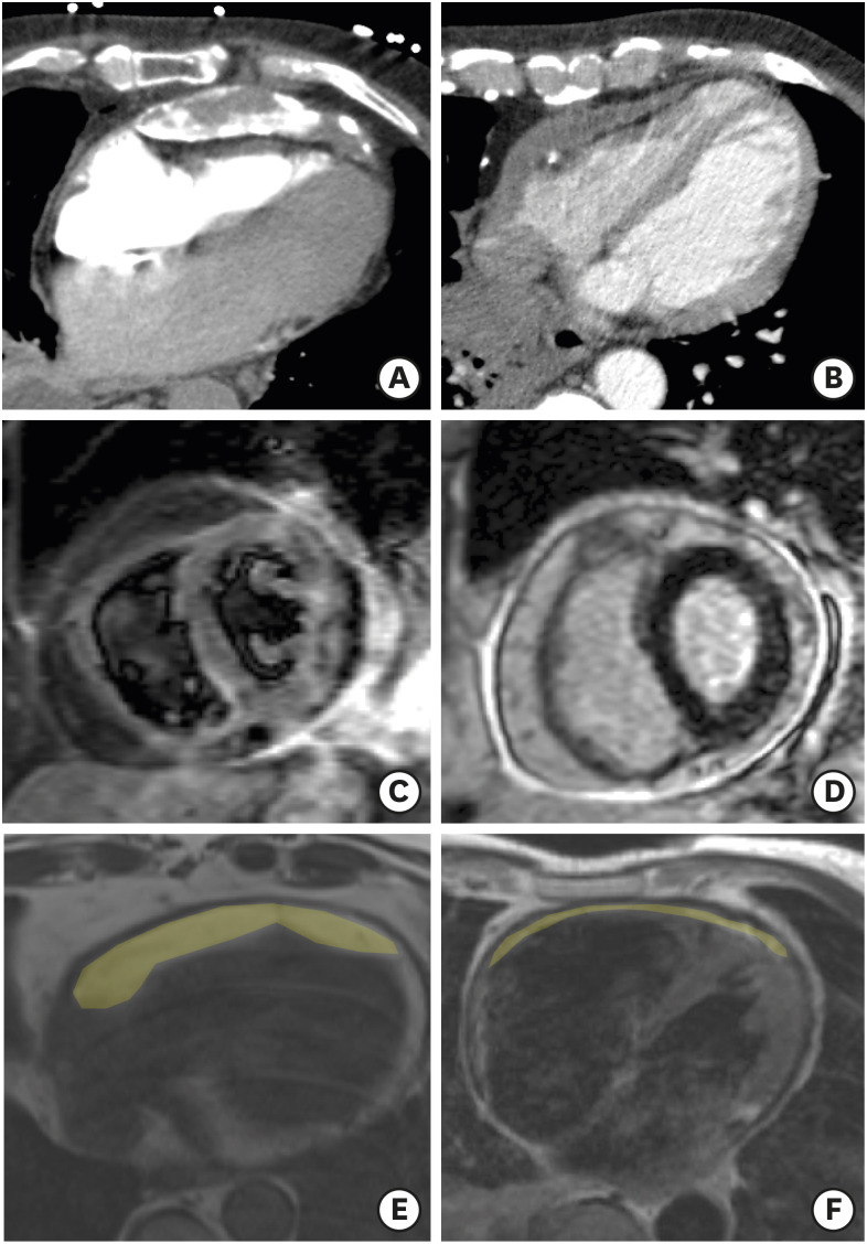
Figure 1. Representative examples of cardiac computed tomography images with (A) thickened and calcified pericardium; (B) thickened but not calcified pericardium. Representative examples of CMR images showing positive (C) pericardial T2-STIR images and (D) pericardial late-gadolinium enhancement. Trans-axial slices from turbo spin echo CMR showing thick (E) compared to thin (F) epicardial fat pad (shaded in yellow).
CMR: cardiac magnetic resonance, echo: echocardiographic, T2-STIR: T2 weighted short tau inversion recovery.

