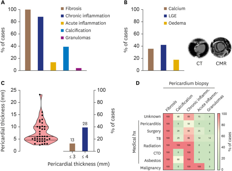
Figure 2. (A) Histological findings of pericardial biopsy and (B) pericardial imaging by CT and CMR in 53 patients with constrictive pericarditis undergoing pericardiectomy. (C) Distribution of pericardial thickness by CT or CMR, and percentage of CP patients with a thickened pericardium. (D) Heatmap for the relationship between cause-specific CP and the prevalence of histological findings.
CMR: cardiac magnetic resonance, CP: constrictive pericarditis, CT: computed tomography, CTD: connective tissue disorder, LGE: late gadolinium enhancement, TB: tubercolosis.

