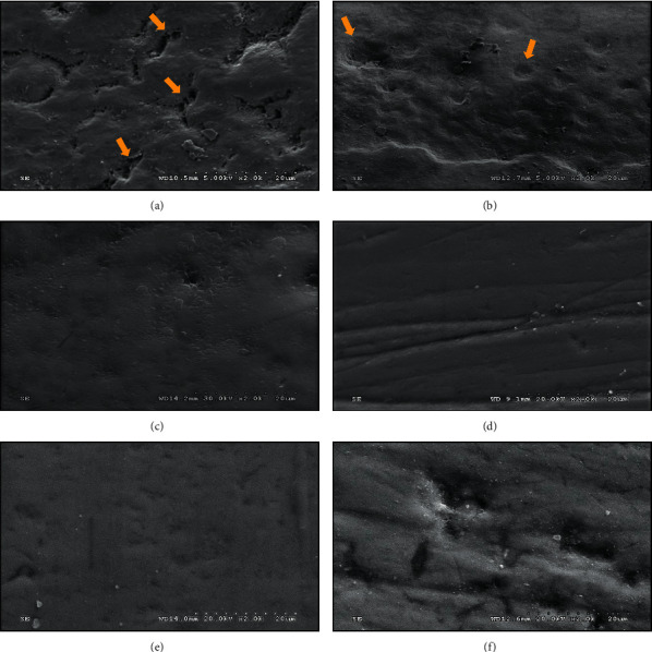
Figure 4.

The scanning electron photomicrograph (SEM, x2K) of the demineralized surface of enamel, showing outlines of enamel prism/rod with the remnant of interprismatic enamel (yellow arrow) (a), followed by remineralization with fluoride varnish, showing incomplete filling of the porosities (yellow arrow) (b), nano-hydroxyapatite toothpaste (c), 20% nano-hydroxyapatite gel (d), 30% nano-hydroxyapatite gel (e) compared to no treatment group (f).
