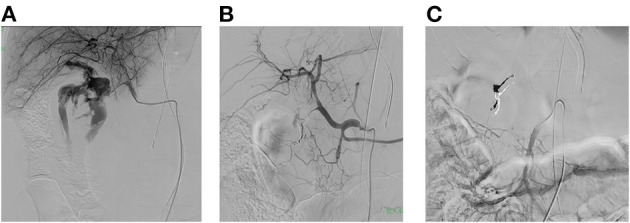
Figure 1.

(A) Imaging is performed via the hepatic artery with the REBOA balloon inflated. A large volume of extravasation from the gastroduodenal artery is seen in the duodenum. (B) After completion of the embolization procedure, imaging is performed via the celiac artery with the REBOA balloon deflated. The gastroduodenal artery has been embolized with coils, and the extravasation has almost completely disappeared. (C) After completion of the embolization procedure, imaging is performed via the superior mesenteric artery with the REBOA balloon deflated. The ischemic area associated with embolization was small, and there were no significant complications.
