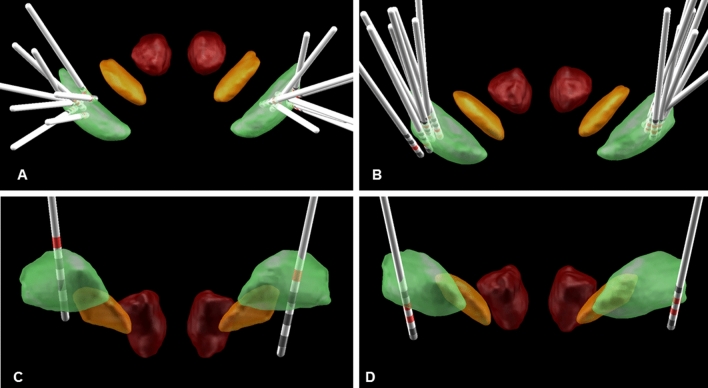
Fig. 1.
a, b 3D group visualization of pallidal electrode locations in MNI space highlighting the active contacts (red) in 17 out of 19 patients using the atlas as described in Ewert et al. [25]. Anatomical structures as defined: internal pallidum (green), subthalamic nucleus (orange), and red nucleus (red). a Electrode localization of the patients with generalized dystonia (n = 8; patients 1 and 9 had to be excluded due to missing preoperative MRI data). b Localization analysis of the CD/SD patients (n = 9). Electrode localization of patient 2 (c) and 4 (d) with poor DBS effects. Electrodes are localized within the Gpi. However, clinical choice of active contacts does not perfectly match the visualized boundaries of Gpi. Consequent stimulation adjustments have been successfully initiated (data not shown, here)

