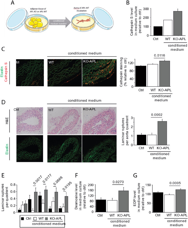
Figure 6.
Effects of conditioned medium obtained from visceral adipose tissue of Apelin-knockout mice (KO-APL mice, grey bars, n = 4) and wild-type mice (WT, white bar, n = 4) on elastic fiber fragmentation of WT aorta. (A) Schematic of the protocol of conditioned medium, applied to the aortas of WT mice. The control condition (Crtl, black bar) corresponded to a medium that had not been in contact with adipocytes. Image was modified from Servier Medical Art, licensed under a Creative Common Attribution 3.0 Generic License. http://smart.servier.com/. (B) Identification of the activity of cathepsin S in the media after incubation of visceral adipocytes from APL-KO or WT mice but before the incubation with WT aorta. (C) Anti-cathepsin S immunostaining in the aorta after 3 days of incubation with the conditioned medium. (D) Histology (hematoxylin–eosin [H&E]) and autofluorescence of aorta elastin after 3 days of incubation with the conditioned medium. (E) Number of laminar ruptures per quadrant after 3 days of incubation with the conditioned medium. (F) Desmosine level found in culture medium, after 3 days of incubation with the conditioned medium (G) Elastin-derived peptide (EDP) level found in culture medium, after 3 days of incubation with the conditioned medium.

