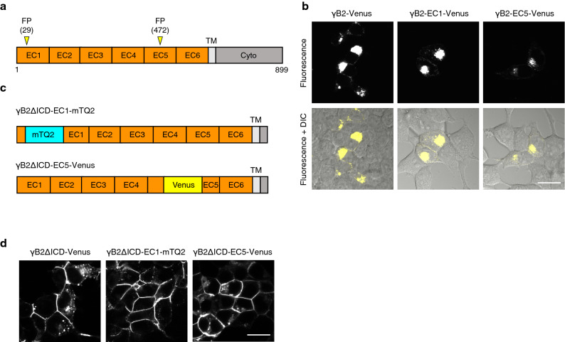
Figure 1.
Molecular design and cellular localization of FP-inserted γB2. (a) Schematics of full-length protocadherin-γB2 (γB2). γB2 is represented as serially repeated extracellular domains (EC domains), a transmembrane region (TM), and a cytoplasmic region (Cyto). The insertion positions (amino acid position 29th and 472nd residues in the matured form) of fluorescent protein (FP) are indicated by yellow arrowheads. (b) The localization of Venus-inserted γB2 constructs in HEK293T cells. HEK293T cells expressing the indicated constructs were observed using a confocal microscope. Fluorescence images (upper) and merged fluorescence and differential interference contrast (DIC) images (lower) are shown. Scale bar, 20 μm. (c) Schematics of FP-inserted γB2ΔICDs. In γB2ΔICD-EC1-mTQ2 and γB2ΔICD-EC5-Venus, mTurquoise2 (mTQ2) and Venus are inserted in γB2ΔICD at the positions as indicated in (a). ΔICD denotes deletion of an intracellular domain (ICD), which leads to efficient localization of γB2 at the plasma membrane. (d) The localization of FP-inserted γB2ΔICDs in HEK293T cells. HEK293T cells expressing the indicated constructs were observed using a confocal microscope. Scale bar, 20 μm.

