Figure 1.
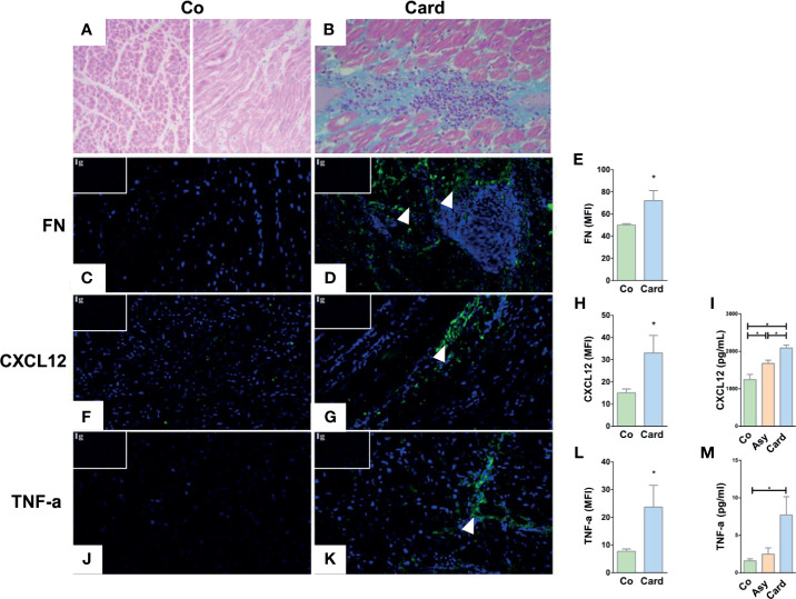
Histological and immunofluorescence analyses of the cardiac tissue of Control subjects and T. cruzi–infected patients with chronic myocarditis. Hematoxylin and eosin staining of healthy myocardium from Control individuals (A) and from Cardiac patients (B) showing the typical diffuse inflammatory infiltrate of T. cruzi chronic infection associated with the inflammatory cell influx. Immunofluorescence images reveals an increased fibronectin (FN) deposition (fibronectin in green, cell nuclei in blue) in the heart sections from Cardiac patients compared to Controls (C, D). The mean fluorescence intensity (MFI) of fibronectin was also increased in the Cardiac group (E). CXCL12 immunolabeling (CXCL12 in green, cell nuclei in blue) showed an augmented expression (F, G) and MFI (H) in Cardiac patients compared to Controls. Systemic amounts of CXCL12 are also enhanced in T. cruzi–infected individuals, despite being Asymptomatic or Cardiac (I). Immunofluorescence images of heart sections from Cardiac patients showing an increased TNF-α expression (TNF-α in green, cell nuclei in blue) (J, K) and MFI (L) compared to Controls. Systemic amounts of TNF-α are also enhanced in Cardiac individuals (M). Isotype controls are shown in the upper left corner of each image. In all cases, MFI was evaluated by measuring five images/heart per study group (Control, n = 3 and Cardiac, n = 3). *p < 0,05 versus Controls. Mann-Whitney U test was used for statistical analyses. Co, Control; Asy, Asymptomatic; Card, Cardiac.

