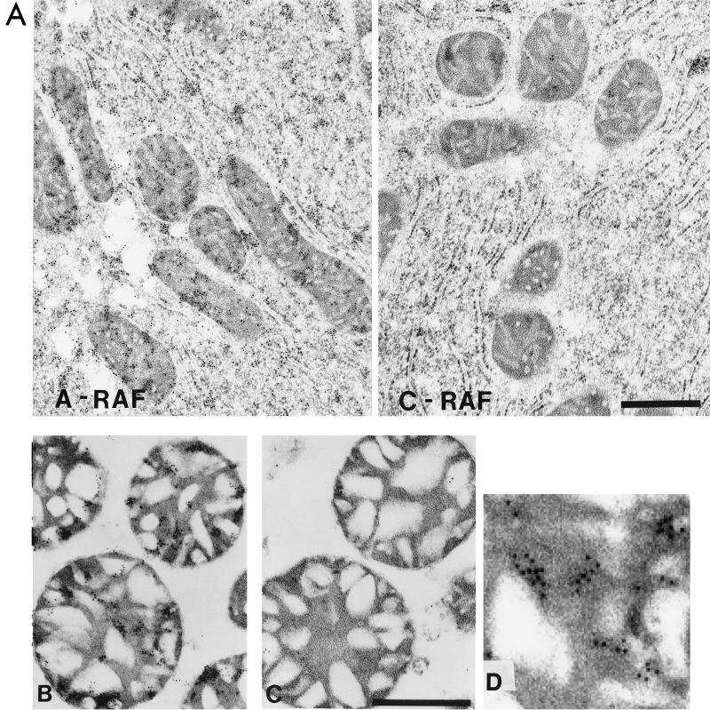
FIG. 6.
Transmission electron micrographs of thin sections of mitochondrial preparations. (A) Rat liver was treated with either A-RAF or C-RAF antibody and stained with gold as described below for mitochondrial sections. The bar equals 1 μm. (B and C) Purified mitochondrial samples were fixed for immunogold labeling with 4% paraformaldehyde-0.1% glutaraldehyde in 0.1 M sodium cacodylate buffer, dehydrated through a graded series of ethanol, and embedded in LR White resin (London Resin Company). Sections of embedded mitochondria were blocked with TBS containing first 50 mM glycine and then containing 5% BSA and 5% goat serum. After overnight incubation in TBS with 1% BSA containing different primary antibody dilutions of anti-A-RAF polyclonal antibody (Santa Cruz Biotechnologies catalog no. sc-408) (B) or anti-C-RAF polyclonal antibody (Santa Cruz Biotechnologies catalog no. sc-227) (C), sections were washed six times with 1× TBS–1% BSA and incubated for 2 h with a 1:10 dilution of goat anti-rabbit antibody conjugated to 10-nm colloidal gold (Goldmark Biologicals). The bar equals 0.5 μm. (D) Enlargement of a section of panel B showing the gold particles.

