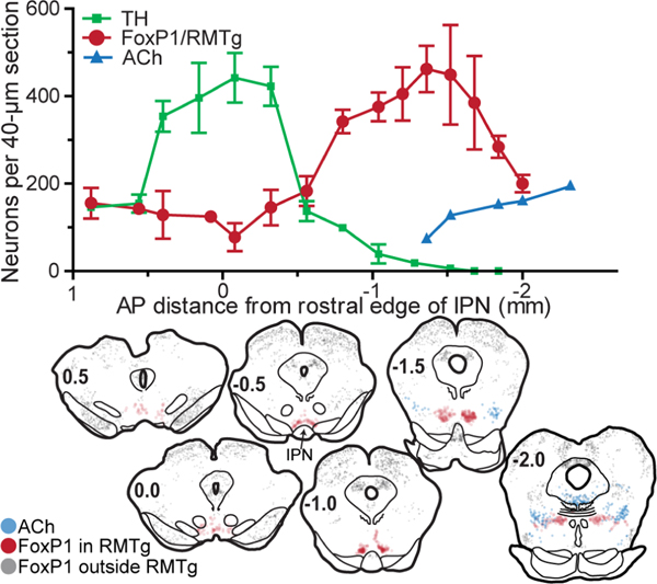
Figure 2.
Distribution of FoxP1/RMTg neurons at various rostro-caudal levels in coronal rat brain sections. (A) Counts of neurons in 40-micron sections plotted versus anterior-posterior distance from rostral edge of IPN. RMTg neurons, as identified by FoxP1 immunostaining, reside caudal to the majority of VTA dopamine neurons, identified via tyrosine hydroxylase (TH), and rostral to many cholinergic neurons of the pedunculopontine nucleus. (B) Coronal rat brain sections show distribution of FoxP1 neurons inside RMTg (red symbols), which are distinct from FoxP1 neurons outside the RMTg (grey symbols). Cholinergic neurons of the PPTg are denoted by blue symbols.

