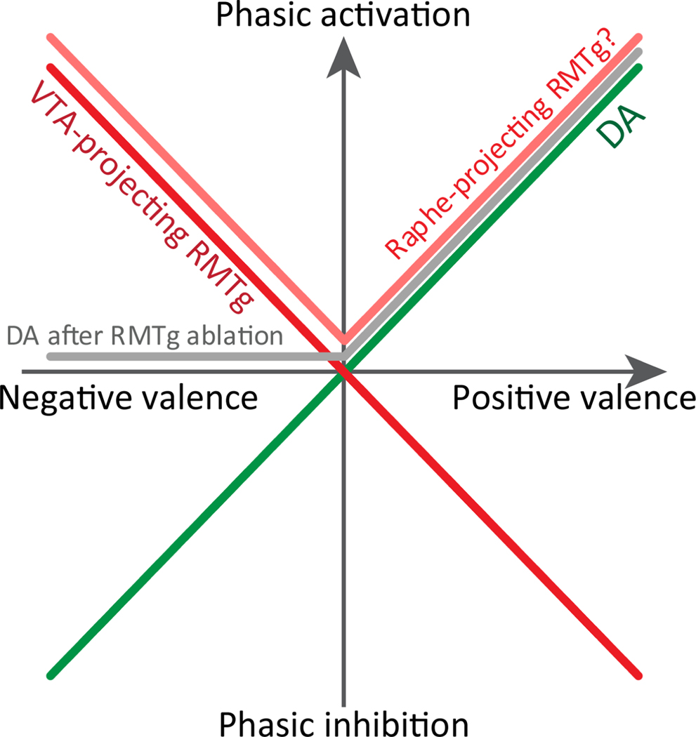
Figure 3.
Simplified depiction of RMTg and DA responses to stimuli of positive and negative valence. Responses of dopamine neurons are shown in both intact rats (green trace) and after ablation of RMTg (grey trace). Responses to RMTg neurons projecting to VTA versus raphe are adapted from (Li et al., 2019a).

