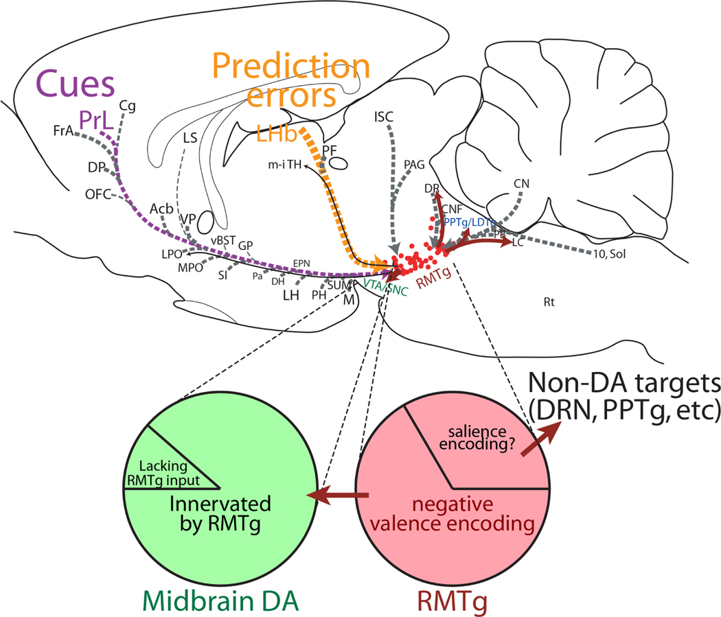
Figure 4.
Sagittal drawing of RMTg efferents (red) and afferents (grey). Afferents with verified electrophysiological influences on RMTg are indicated with colored lines and text: prelimbic cortex (PrL, purple), and lateral habenula (LHb, orange). RMTg and dopamine neuron populations are further dissected in pie chart diagram, showing diversity of neuron subtypes with proportions approximated by areas of pie chart. Roughly one-third of RMTg neurons may project to targets outside the midbrain dopamine system, while a minority of dopamine neurons may lack an RMTg input.

