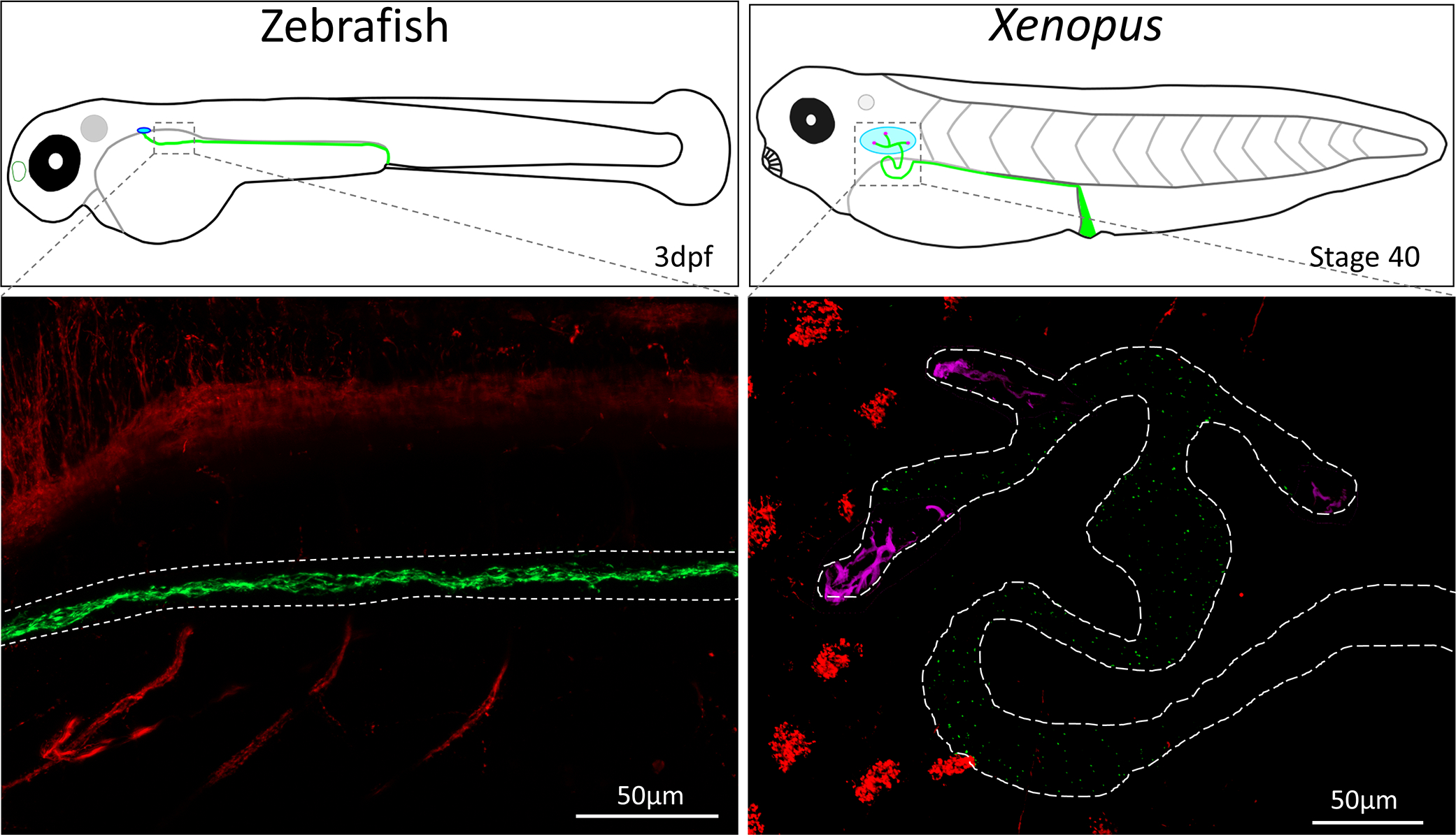
Figure 2:

Confocal images of wholemount zebrafish (3dpf) and Xenopus laevis (Stage 37) kidney cilia. Cilia were stained using an acetylated alpha-tubulin antibody (Sigma T6793) which labels the neurons and cilia. Kidney cilia are pseudocolored in green while neurons and epithelial cilia are pseudocolored in red. The zebrafish and Xenopus kidney are outlined in white dashed lines, and motile multiciliated cells in the kidney are pseudocolored in magenta. Images were taken on a Zeiss LSM800 confocal microscope.
