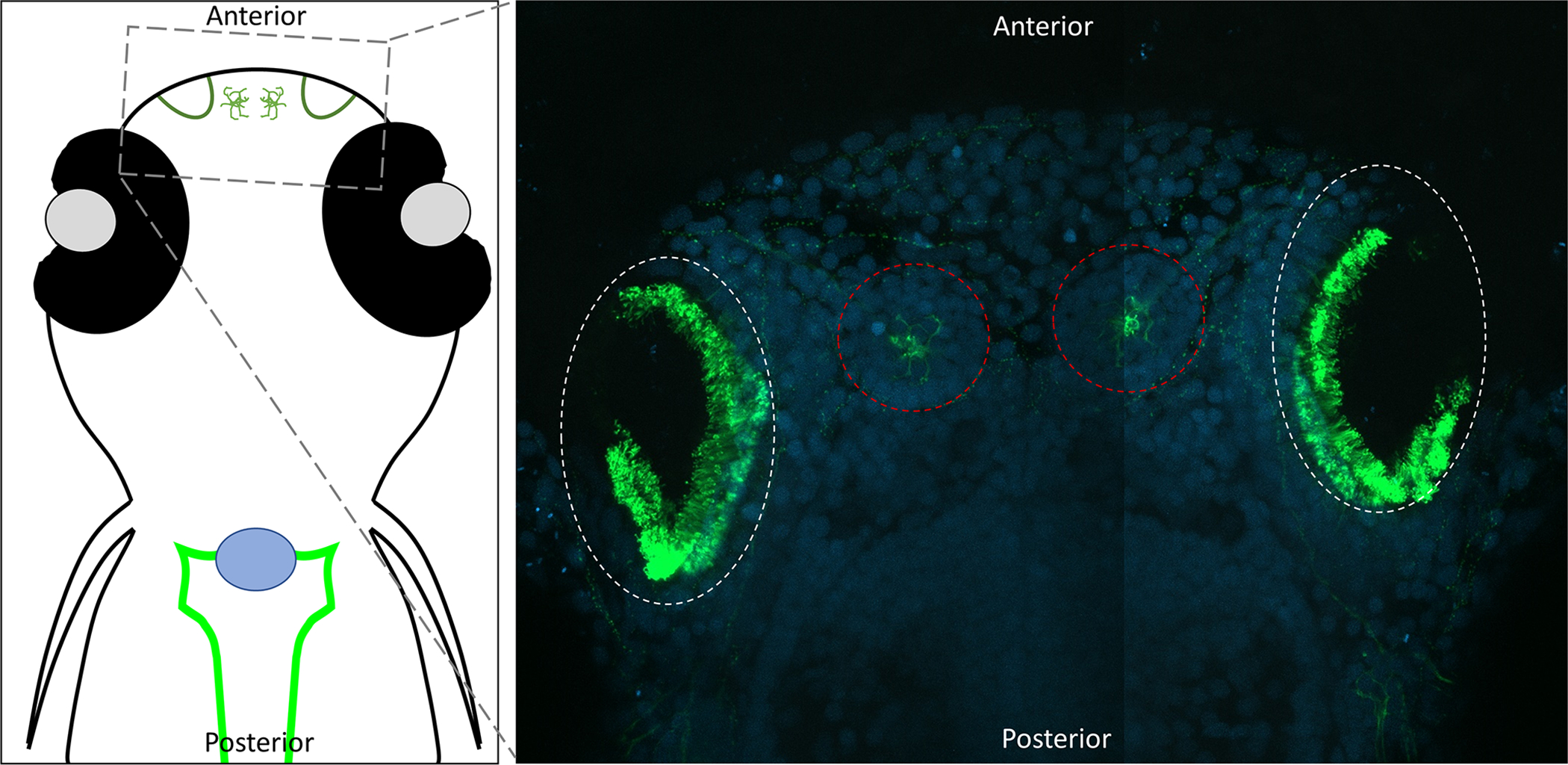
Figure 4:

Confocal images of the motile cilia lining the zebrafish nasal (olfactory) pit. Dorsal view of 8dpf zebrafish embryos with head towards the top of the image. Embryos were fixed and stained with acetylated alpha-tubulin (Green) (Sigma T6793) and DAPI (Blue). Acetylated tubulin labels both the cilia and neurons. Nasal pits are circled in white, and neural mast cells are circled in red.
