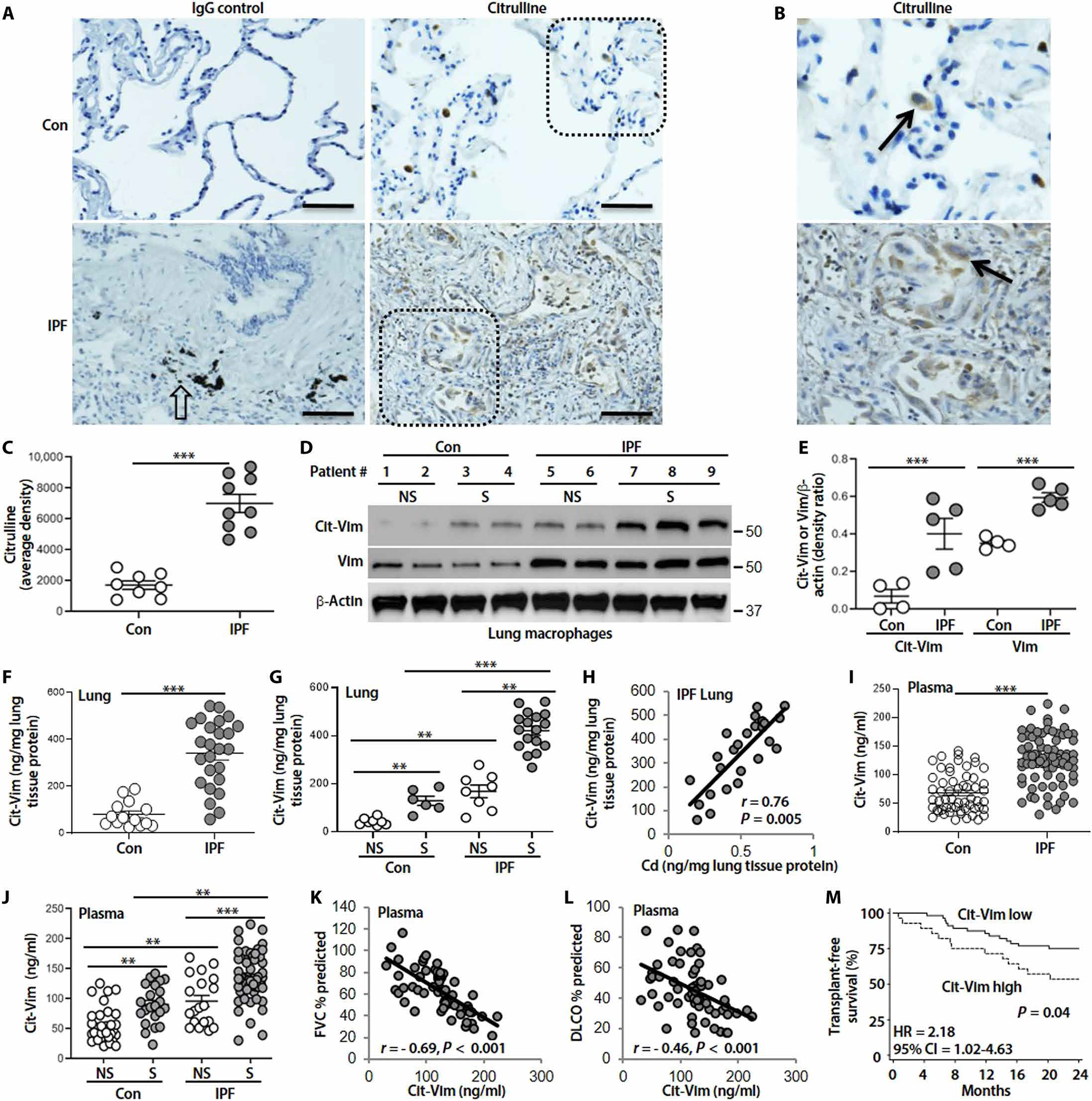
Fig. 2. Cd positively correlates with Cit-Vim, and Cit-Vim correlates with lung function parameters and transplant-free survival.

(A) Representative human lung histology with citrulline staining (arrows: stained brown) from control (n = 8) and subjects with IPF (n = 9). (B) Higher magnification of inset from (A). Open arrow indicates CB. Scale bars, 100 μm. (C) DAB staining density from (A) was quantified. (D) Immunoblot analysis of Cit-Vim and Vim in lung macrophage. (E) Quantification of Cit-Vim and Vim expression from (D). (F) Cit-Vim amounts by ELISA in lung tissues and (G) analyzed by smoking status. (H) Correlation analysis of Cd with Cit-Vim in lung tissue from subjects with IPF (n = 25). (I) Cit-Vim amounts by ELISA in plasma and (J) analyzed by smoking status. Correlation analysis of plasma Cit-Vim with predicted percentage of (K) FVC and (L) DLCO. P and r values were determined using Spearman rank correlations. (M) Transplant-free survival analysis during the next 2 years between the subjects with higher Cit-Vim and subjects with lower Cit-Vim. HR and CI values were established by Cox proportional hazard regression. **P < 0.01 and ***P < 0.001 using two-tailed t test for (C) and (F) and one-way ANOVA followed by Tukey’s post hoc analysis for (E), (G), and (J).
