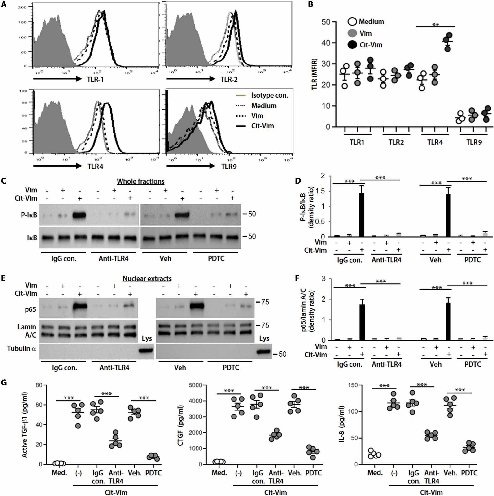
Fig. 5. Cit-Vim exposure induces TLR4/NF-κB-mediated cytokine/chemokine production.

(A) Representative histograms on flow cytometry showing surface expression of TLR1, TLR2, TLR4, and intracellular TLR9. (B) Quantification TLR expression using the MFIR as shown in Fig. 4D. Lung fibroblasts isolated from normal subjects were incubated for 48 hours, and each dot represents each subject. (C) Immunoblot analyses of p-IκB and IκB expression in whole fractions. (D) Quantification of p-IκB expression from (C) (n = 3). (E) p65 and lamin A/C expression in nuclear extracts. (F) Quantification of p65 expression from (E) (n = 3). Lung fibroblasts were preincubated with IgG control, anti-TLR4 antibody (10 μg/ml), vehicle, or NF-κB inhibitor (pyrrolidine dithiocarbamate, PDTC, 100 μM) for 1 hour and then stimulated with Vim or Cit-Vim (2 μg/ml) for 2 hours. Lamin A/C was used as a nuclear protein loading control, and α-tubulin was used as a cytoplasm protein control. (G) Cytokine/chemokine concentrations by ELISA in fibroblast supernatants stimulated with Cit-Vim for 24 hours in the presence of IgG control, anti-TLR4 antibody, vehicle, or PDTC. **P < 0.01 and ***P < 0.001 using one-way ANOVA followed by Tukey’s post hoc analysis.
