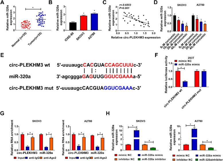
Fig. 4.
Circ-PLEKHM3 sponged miR-320a. A MiR-320a expression was detected in tumor and normal tissues (n = 35). B MiR-320a level was measured in SKOV3, A2780 and IOSE-80 cells. C The linear correlation of miR-320a and circ-PLEKHM3 in ovarian cancer tissues was analyzed. D MiR-320a abundance was examined in SKOV3 and A2780 cells after exposure to various concentrations of curcumin. E The binding site of circ-PLEKHM3 and miR-320a was predicted via starBase. F Luciferase activity was detected in 293 T cells with transfection of circ-PLEKHM3 wt or mut and miR-320a mimic or mimic NC. G Circ-PLEKHM3 and miR-320a abundances were detected after RIP. H MiR-320a abundance was examined in SKOV3 and A2780 cells transfected with mimic NC, miR-320a mimic, inhibitor NC or miR-320a inhibitor. *P < 0.05

