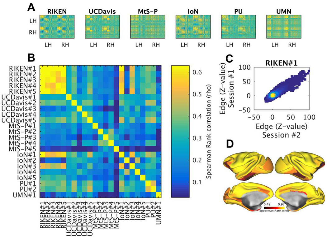
Fig. 8. Reproducibility of resting-state functional connectivity (FC) within and across PRIME-DE macaque imaging sites.

(A) Exemplar FC correlation matrices from six PRIME-DE sites, ordered according to hierarchical clustering (Ward’s method). (B) Comparison between correlation matrices across sites (six) and subjects (total N=23). fMRI timeseries were preprocessed using HCP-NHP pipelines, parcellated using M132 atlas containing 91 parcels per hemisphere (Markov et al., 2014), and then Spearman’s Rank correlation coefficient (rho) between parcellated timeseries was calculated. Comparison of FC was limited to PRIME-DE sites that fulfilled minimum acquisition criteria (high-resolution anatomical image and a B0 field-map). (C) Test-retest (heat) scatter plot of Z-scored FC matrixes (N=1, n=2, RIKEN data). (D) Reproducibility was high (>0.8; rho) in majority of the cortex (>78%), however, areas distant to RF receive channel coils and weaker SNR (i.e. hippocampal complex and ventral visual stream) exhibited lower reproducibility (RIKEN data was acquired using HCP-style protocols). Abbreviations: HCP the human connectome project; UC-Davis University of California, Davis; MtS Mount Sinai-Philips; IoN Institute of Neuroscience; PU Princeton University; UMN University of Minnesota, RH right hemisphere; LH left hemisphere.
