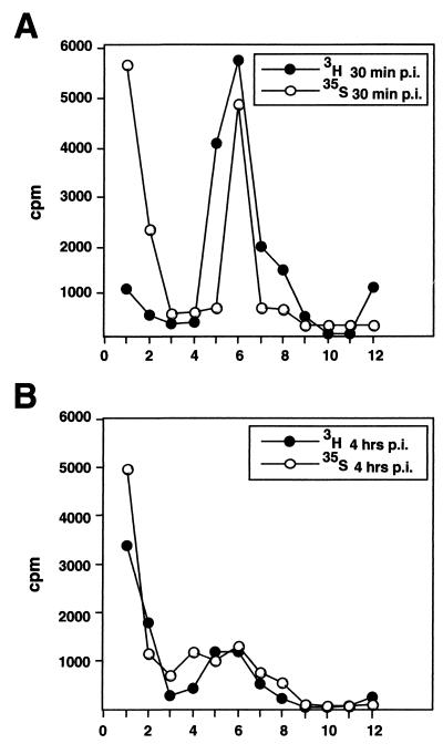
FIG. 2.
Uncoating of HSV-1 in Vero cells. Vero cells were infected with [3H]thymidine- or [35S]methionine-labeled virus at an MOI of 10 PFU/cell. At various time points, the extracellular viruses were removed by proteinase K and the cells were lysed. The postnuclear supernatants were loaded onto a linear sucrose gradient for ultracentrifugation analysis of the internalized capsids. The sedimentation profiles of internalized [3H]thymidine-labeled and [35S]methionine-labeled capsids at 30 min (A) and 4 h (B) p.i. are shown. Sedimentation profiles from one experiment are shown. However, the analysis was repeated three times, and mean values (with standard deviations) of two independent experiments with [3H]thymidine-labeled virus presented as percentages of total counts are shown in Table 1. The x axes indicate fractions.

