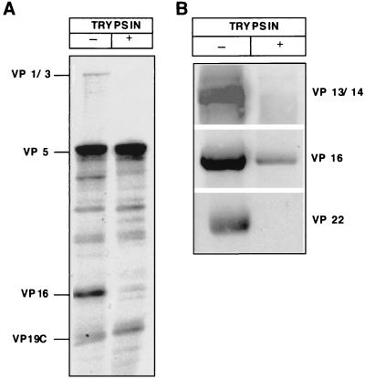
FIG. 3.
Characterization of HSV-1 capsids isolated from virions in vitro. (A) Coomassie blue staining of an SDS–10% PAGE analysis of the capsids. The capsids were isolated as a light-scattering zone in the sucrose gradient (left lane). Capsids were treated with 10 μg of trypsin per ml at 37°C for 5 min (right lane). The HSV-1 proteins identified by molecular weight are indicated on the left. (B) Western blot of the in vitro-isolated capsids (left lane) and capsids treated with 10 μg of trypsin per ml (right lane). The proteins identified with specific antibodies are indicated on the right.

