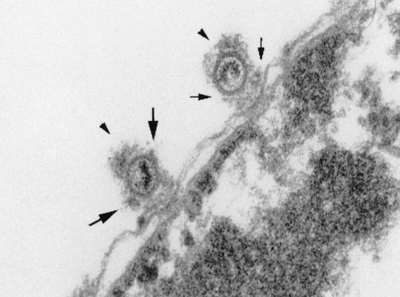
FIG. 5.
Preferred binding of HSV-1 capsids to NPCs. An electron micrograph of capsids (arrowheads) binding to rat liver nuclei in vitro is shown. Small arrows indicate the cytoplasmic fibrils emanating from the NPC, and large arrows show the additional electron-dense material at the capsid vertices.

