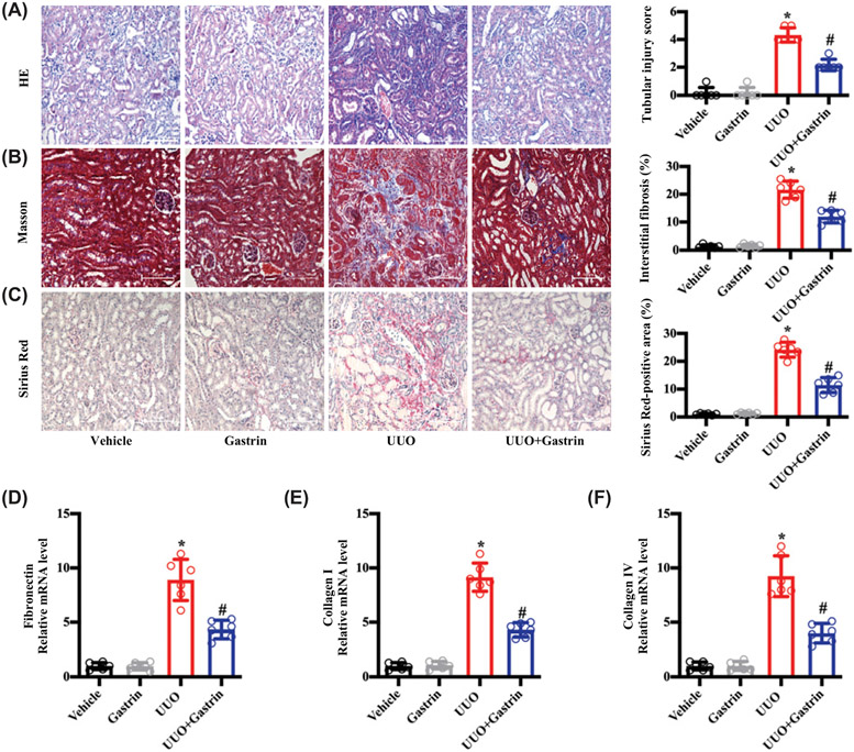
Figure 3. Gastrin attenuates renal fibrosis caused by UUO.
Mice with UUO or sham-operated mice were infused subcutaneously, via an osmotic minipump with saline vehicle (control, 2.64 μl/d) or gastrin (120 μg/kg body weight /d x 7 d). The samples were collected at the end of 7 d. (A) Representative images of H&E staining and quantitative analysis of renal tubular injury. n=6; *P<0.05 vs. Vehicle treatment; #P<0.05 vs. Ang II treatment. (B) Representative images of Masson’s trichrome staining and quantitative analysis of renal interstitial fibrosis. n=6; *P<0.05 vs. Vehicle treatment; #P<0.05 vs. Ang II treatment. (C) Representative images of Sirius Red staining and quantitative analysis of Sirius Red-positive areas showing renal collagen deposition. n=6; *P<0.05 vs. Vehicle treatment; #P<0.05 vs. Ang II treatment. (D–F) Fibrosis-related mRNA levels in the kidney: fibronectin (D); collagen I (E); and collagen IV (F). n=6; *P<0.05 vs. Vehicle treatment; #P<0.05 vs. Ang II treatment.

