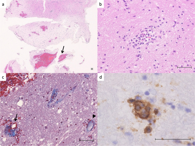
Fig. 2.
Histology of the hematoma wall and cerebral tissue. a H&E stain shows scattered petechial hemorrhages and perivascular hematoma (arrows) in the cerebral cortex. b Typical angiocentric neutrophilic infiltration with few cell fragments and mild extravasation of red blood cells. There is no necrosis in the surrounding brain tissue. c Masson Trichrome stain shows that the small blood vessel in the hemorrhage area (arrow) is disrupted and has collagen fiber tears and extravasation of red blood cells compared to the open, uninvolved small blood vessel (arrowhead). d High power view of the arrow area in Fig. 2c. CD31 immunostaining shows that the nuclei of vascular endothelial cells are enlarged and disorganized. Scale bars are 50 μm

