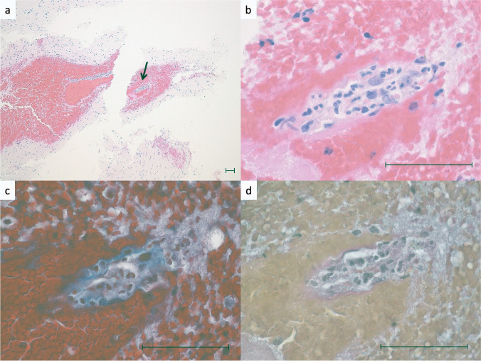
Fig. 3.
Histological features of vasculitis. a Middle power view of the perivascular hematoma. b High power view of the arrow area in Fig. 3a. The small blood vessel is compressed and occluded by the perivascular hematoma. Neutrophilic infiltration is observed in the vessel wall, and endothelial cell enlargement and fibrin deposition are shown. c Same area as in Fig. 3b. Masson Trichrome stain shows tearing and obscuration of collagen fibers in the wall of the small vessel. d Same area as in Fig. 3b. Elastica van Gieson staining shows elastic fibers and smooth muscle are absent in the vessel wall. Scale bars are 50 μm

