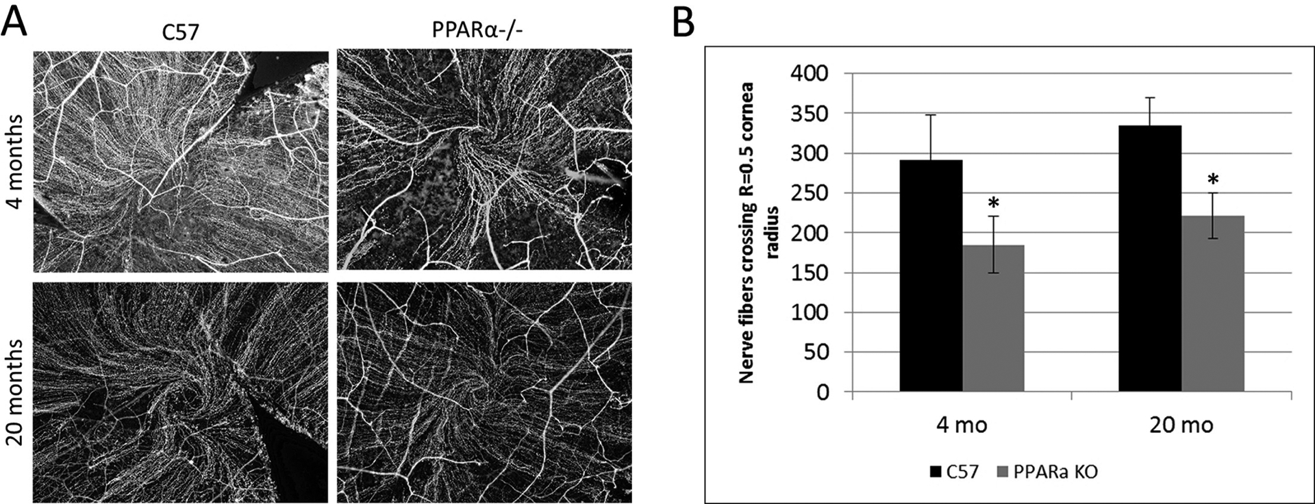
Figure 6:

Decreased corneal nerve fibers in PPARα−/− (KO) mice compared to C57Bl/6J (WT). A) Corneal nerve fibers immuno-labeled with an antibody for beta-III neuron-specific tubulin. WT and KO nerve fiber densities were compared at 4 and 20 months of age by counting nerve fibers crossing 0.5 times the cornea radius. B) Maximum central corneal nerve fiber count: mean ± SEM; *P≤0.05, rel. to WT; 4 months N(WT)=7, N(KO)=6; 20 months N(WT)=10, N(KO)=9. Modified from Matlock et al. (2020).
