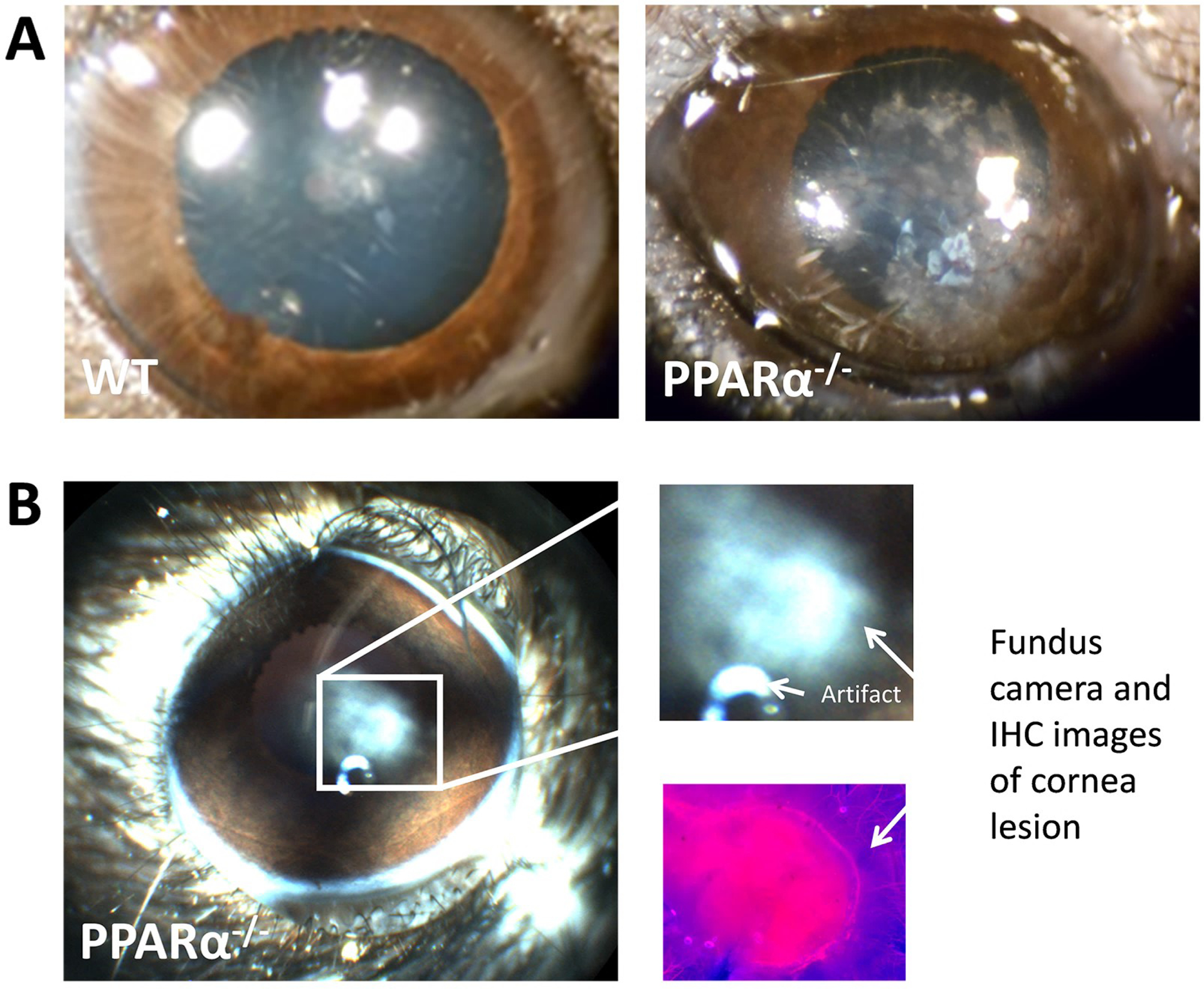
Figure 7:

Cornea lesion in the PPARα KO mouse. A) Photographs of wild-type mouse cornea with normal opacity (left) and large PPARα KO mouse cornea lesion at 24 months of age. B) Fundus camera image of small PPARα KO cornea lesion in 24-month-old mouse (left), explode view (right), and neuron-specific beta-III tubulin immuno-labeling from the same lesion (lower right). Modified from Matlock et al. (2020).
