FIG 4.

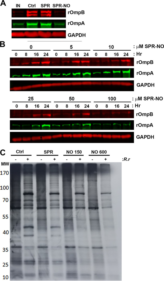
NO inhibits protein synthesis of intracellular R. rickettsii. (A) Vero cells were infected with R. rickettsii for 2 h, and select samples were collected immediately (IN) or were treated with 150 μM SPR or SPR-NO and then collected 24 h later. Samples were processed for Western blotting with anti-rOmpA, anti-rOmpB, and anti-GAPDH antibodies (representative blot, n = 3). (B) Vero cells were infected with R. rickettsii for 2 h and treated with increasing concentrations of SPR-NO. After 0, 8, 16, and 24 h of treatment, samples were collected for Western blotting. Anti-rOmpA, anti-rOmpB, and anti-GAPDH antibodies were used (representative images, n = 5). (C) Vero cells were infected with R. rickettsii for 24 h, treated with SPR or SPR-NO for 3 h, and then labeled with [35S]Met for 3 h. Samples were scraped into Laemmli sample buffer, boiled, and separated by SDS-PAGE, gels were dried and exposed to film, and autoradiograms were visualized (representative autoradiogram, n = 3).
