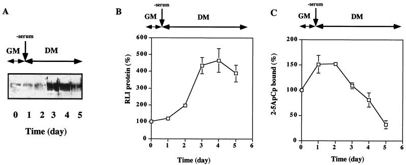
FIG. 1.
Increases in the RLI and RNase L proteins during the differentiation of C2 myoblasts into myotubes. (A) C2 cells were grown in GM and then shifted to DM on day 1. At the indicated times, cells were harvested and lysed as described in Materials and Methods. Total protein samples (100 μg) were analyzed for RLI protein by Western blotting with a polyclonal RLI antiserum. (B) Densitometric analysis of the gel shown in panel A. A value of 100% corresponds to the amount of RLI protein in proliferating myoblasts at day 0. Error bars refer to the standard deviation obtained in three independent experiments. (C) C2 cells were grown in GM and then shifted to DM on day 1. At the indicated times, cells were harvested and lysed. Proteins (600 μg) were incubated with radiolabeled 2-5A4-3′-[32P]pCp (2-5ApCp) in a radiobinding assay; 100% corresponds to the amount of 2-5ApCp bound to RNase L in proliferating myoblasts. Error bars refer to the standard deviation obtained in three independent experiments.

