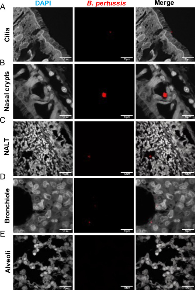
FIG 8.

Immunofluorescence (IF) staining of B. pertussis localization in the respiratory tract. B. pertussis was labeled using a polyclonal antibody to FHA and counter-tagged with a fluorescently conjugated antibody (Texas Red). Sections were counterstained with DAPI. (A to C) Representative images of B. pertussis in the nasal cavity over the course of infection. B. pertussis was found captured in the cilia of the nasal cavity, as well as of the NALT. (D and E) Representative images of the bronchioles and alveoli of infected rats. B. pertussis was found localized in the bronchioles over the course of infection and absent in the alveoli.
