Figure 2.
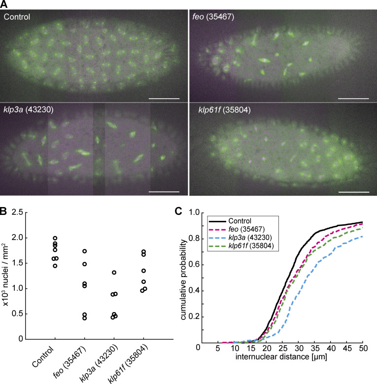
Knockdown of feo, klp3a, or klp61f by RNAi leads to defective nuclear delivery to the embryo cortex. (A) Maximum-intensity projections from 3D time-lapse videos of embryos expressing RNAi against mCherry, feo, klp3a, or klp61f and expressing Jupiter::GFP (green) marking microtubules and H2Av::RFP (magenta) marking chromatin. Knockdown embryos showed irregular nuclear distribution during the first interphase occurring at the cortex as compared with the regular nuclear distribution in control embryos (mCherry). Scale bar, 50 µm. (B) A quantification of the number of nuclei per square millimeter showed a higher degree of variation between the six embryos knocked down for either of the three genes, feo (35467), klp3a (43230), or klp61f (35804), as compared with control embryos. In all cases, the density decreased on average. Each data point represents one embryo. (C) The cumulative probability function of the internuclear distance between first-order neighbors in embryos inhibited for Feo, Klp3A, or Klp61F expression showed a flatter distribution and a median shifted to higher internuclear distance. Thus, the number of nuclei at the cortex was smaller with broader distribution, indicating greater irregularity and larger unoccupied areas with respect to the control. n = 7 (control), n = 6 (RNAi lines). Refer to Table 1, Fig. S2, and Video 5.

