Figure 5.
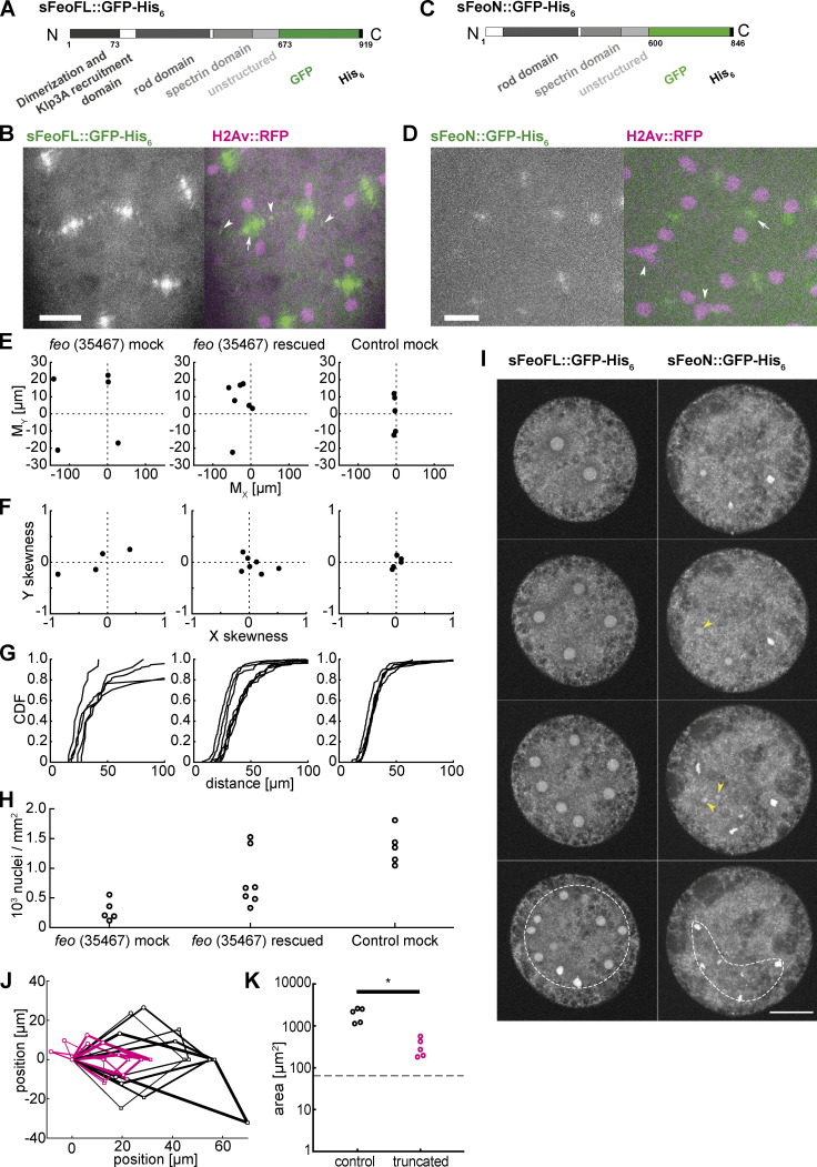
Purified Feo protein rescues nuclear separation in feo RNAi embryos, and an N-terminally truncated Feo abolishes nuclear separation. (A) Scheme of the synthesized full-length Feo protein fusion construct containing a C-terminal GFP. The domains were determined based on sequence similarity from reported domains of the human construct. The N-terminal end induces dimerization and binds Klp3A, and the spectrin domain binds to microtubule lattice. (B) Fluorescence image of the GFP-tagged full-length Feo protein in a (control) blastoderm embryo in telophase after protein injection. The GFP signal alone (left) is shown merged with H2Av::RFP in magenta (right). sFeoFL::GFP-His6 localized correctly, under cell cycle control, at the spindle midzone (arrow) and between daughter nuclei (arrowhead) as observed in the transgenic overexpression fly line shown in Fig. 1. Scale bar, 10 µm. Refer to Video 8. (C) Scheme of a truncated Feo construct lacking the first 73 amino acids of the putative dimerization and Klp3A recruiting domain, fused to a C-terminal GFP. (D) Fluorescence image of the GFP-tagged truncated Feo protein in a (control) blastoderm embryo after protein injection. The GFP signal alone (left) is shown merged with H2Av::RFP in magenta (right). A faint GFP signal localized at the spindle midzone (arrow) in the second division after injection (Fig. S5 C). Nuclear separation defects manifest as neighboring nuclei touching or fusing after division (arrowheads). Scale bar, 10 µm. Refer to Video 9. (E) Plot of the 2D centroid vector (MX,MY) of all cortical nuclei relative to the embryo center for Feo RNAi embryos either mock injected (left; n = 5) or injected with sFeoFL::GFP-His6 protein (middle; n = 7), compared with mock injected control (mCherry) RNAi embryos (n = 5). The x axis designates the anterior-posterior axis, and the y axis is the dorsoventral axis of the embryo. Deviations from zero mark acentric delivery of nuclei to the cortex. Along the anterior-posterior axis, the injection of Feo full-length protein in Feo RNAi embryos partially rescued centering (middle); mock-injected Feo RNAi embryos had anatomically eccentric nuclei (left); and mock-injected control (mCherry) RNAi embryos exhibited strong centering. (F) Skewness plot of the positional distribution of all nuclei along the anterior-posterior (x) and dorsoventral (y) axes for the same conditions as in E. The asymmetric distribution in mock-injected Feo RNAi embryos (left) is partially rescued by Feo protein injection (middle), while mock-injected control embryos show little asymmetry. (G) Cumulative distribution plot of the first-order neighbor distance between nuclei, for the same conditions as in E and F. The irregular internuclear distances in mock-injected Feo RNAi embryos (left) are rescued to a considerable extent after full-length protein injection (middle), while mock-injected control (mCherry) RNAi embryos exhibit uniform internuclear distances (right). CDF, cumulative distribution function. (H) The low nuclear density arriving at the cortex in mock-injected Feo RNAi embryos is partially rescued when full-length Feo protein is injected in preblastoderm Feo RNAi embryos. (I) Addition of full-length GFP-tagged Feo protein to embryo explants supports normal nuclear division and regular distribution within the explant space (left, white circle), while addition of truncated Feo protein reduces nuclear separation (arrowheads), causing occasional spindle fusion, and abolishes nuclear distribution (dashed envelope). Scale bar, 30 µm. (J) Overlay of aligned quadrilaterals describing the nuclear separation after division in explants, as described in Fig. 4. Explants were generated from wild-type embryos and had ample space for the first few divisions. Experiments involving addition of full-length GFP-tagged Feo protein to the explant are in black (n = 5); experiments involving addition of N-terminally truncated, GFP-tagged Feo protein are shown in magenta (n = 5). (K) The truncated Feo protein significantly reduced nuclear separation, as measured by the area of quadrilaterals shown in J, as compared with the full-length protein construct (black).

