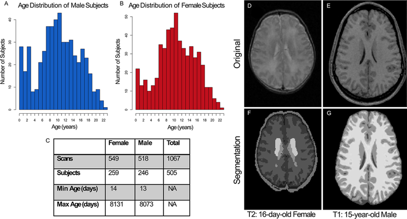
FIG. 1.

Data from each age group from 13 days old to 22 years old were included in the cohort for both males (A) and females (B). The cross-sequential study included 505 total subjects (C), with most having between 2 and 3 longitudinal visits (separated by 2 years). For this study, only scans from 13-day-old to 18-year-old subjects were included to focus on the pediatric age range. Two different algorithms for volume quantification were used, one for the neonates and one for the older cohort. As an example, a T2-weighted MRI scan (D) from a 16-day-old female is shown. This scan was processed through the dHCP pipeline to produce a segmentation (F) including gray matter (found by adding the deep gray matter and cortical gray matter), CSF, and white matter. A T1-weighted MRI scan (E) from an older patient (15-year-old male) is shown. This scan was processed using the CAT12 pipeline within SPM12 to produce the segmentation (G) including gray matter, CSF, and white matter.
