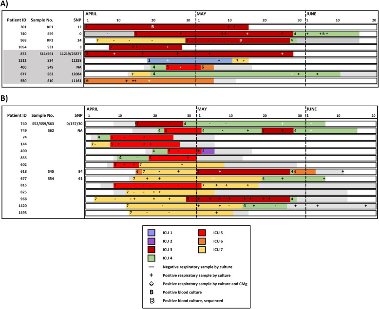
Fig. 2.
Identification of MDR K. pneumoniae and C. striatum outbreaks across the ICU network based on combined epidemiological and CMg analysis. Overlapping ward stays for patients involved in putative outbreaks of A) MDR K. pneumoniae and B)
C. striatum. Each row represents a unique patient. Patients are ordered by ward of first positive (ascending) and then by patient ID (ascending). The horizontal axis shows the ward stays from April 1 to June 20. Non-ITU wards are coloured in grey. ITU wards are labelled 1–10 represented by a unique colour. Periods outside the hospital are represented in white. MDR-K. pneumoniae or C. striatum positive and negative respiratory samples by culture are marked as (+) or (-), respectively. Additionally, positive respiratory samples by CMg and culture are marked as “ ”. Positive blood cultures are marked as “B” and sequenced blood cultures are marked as “
 ”. Patients with a CMg-aligned sequence have an S number (respiratory sample) or KP number (blood culture) adjacent to their identification number on the left of each bar. The number of SNPs for each CMg sample is also shown on the vertical axis. A) CMg was performed on MDR-K. pneumoniae in respiratory samples from patients 1054 and 301 and bloodstream infection isolates on patients 301 and 968 retrieved from the routine diagnostic laboratory (time point marked as “B”). Possible chain of transmission is from top to bottom. No sequenced patient could link 968 to 1517 or 618 to 740 and so were assumed to be due to cryptic transmission via other non-sequenced patients. B) CMg was performed on C. striatum in respiratory samples from patients 618, 677, 740 and 749. All other patients were linked by epidemiology only

