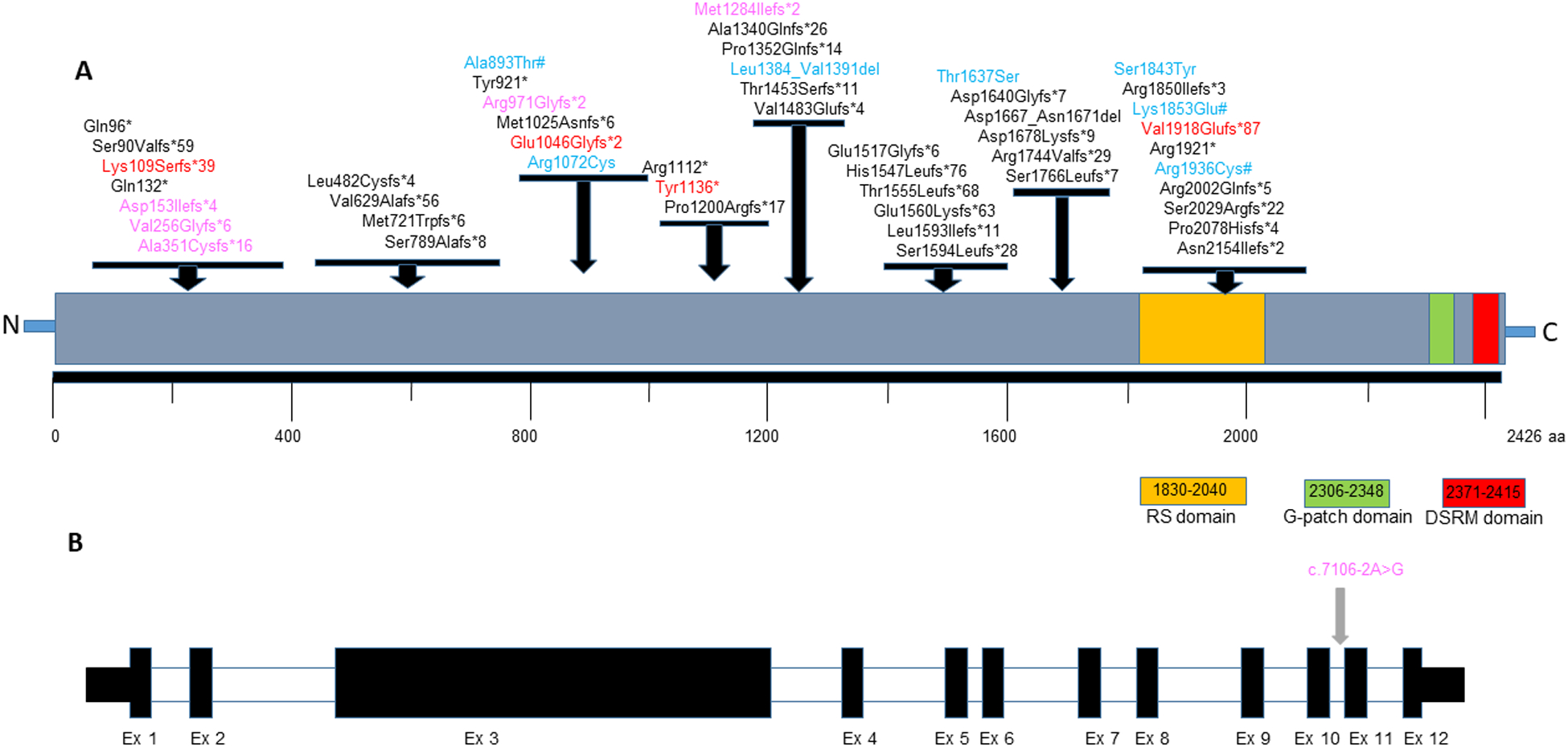
Fig 1:

A. Distribution of variants seen in affected individuals on the SON protein. The LoF variants identified in this study are shown in red, missense & in-frame deletion variants are shown in blue. Predicted loss of function variants from DECIPHER cases are shown in pink and missense changes from DECIPHER cases are shown in blue followed by a #. Other variants reported in literature are shown in black color (NP_620305.3). The location of the functional domains in SON protein were adapted from Hickey et al 2013. B. Schematic showing the exons and the splice variant reported in the SON gene (NM_138927.4).
