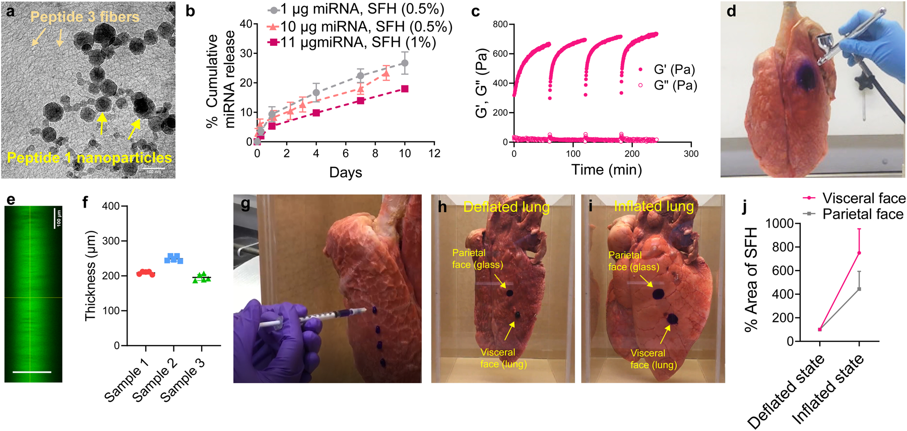
Fig. 4. Formulation and physical characterization of SFH.

a, Cryo-TEM of 1 wt% SFH showing miRNA nanoparticles embedded within the fibrillar gel network formed by self-assembled Peptide 3. Fibers and nanoparticles are labeled. Scale bar 100 nm. Representative of 2 independent experiments. Uncropped micrograph (Figure S26b) b, miRNA particle release from 0.5 and 1 wt% SFH over time. Data = mean ± SD, n = 3 independent samples, representative of 3 independent experiments. c, Time-sweep shear thin-recovery oscillatory rheology of SFH. Storage and loss moduli (G’ and G”) were first monitored for 1 h, after which SFH underwent 3 consecutive shear-thin/recovery cycles. Data representative of 3 independent experiments. d, SFH is sprayed onto the surface of a porcine lung. The material is shear-thinned into microdroplets when sprayed and recovers on the surface of the lung. SFH is doped with Crystal Violet to aid in visualization. e, Confocal microscopic z-stack image of SFH (doped with calcein) sprayed onto a glass surface. f, Quantification of the thickness of sprayed SFH from panel e by measuring the distance (shown with green line) between the bottom of the image (region below the glass slide) and the top of the image (region above the gel). g, SFH is syringe-injected onto the surface of a porcine lung. Supplementary Video 1 shows that SFH adheres and expands with the lung tissue during lung inflation. Supplementary Video 2 shows syringe-based injection and tissue-site localization of SFH. h, SFH syringe-delivered to deflated lung and glass chamber surfaces modeling its application to the visceral and parietal surfaces of the pleural cavity after mesothelioma debulking. i, Spread-fill behavior of SFH during lung re-inflation and tissue contact with the parietal wall; also shown in Supplementary Video 3, 4 and 5. j, quantification of spread area for both the visceral and parietal surface-applied SFH as a function of lung status, Data = mean ± SD, n = 3 independent experiments.
