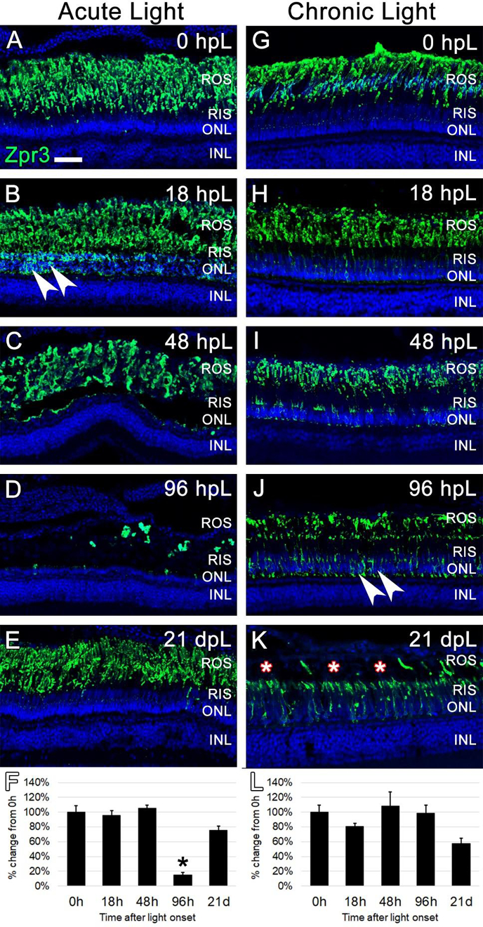
Figure 5. Analysis of rod photoreceptor degeneration/regeneration during acute and chronic light treatments.

In undamaged retinas (A and G), Zpr3 immunocytochemistry (green) labeled rod photoreceptor outer segments (ROS). In the acute group (A-E), Zpr3 additionally localized to the rod inner segments (RIS) and the perinuclear space of rod photoreceptor nuclei in the ONL (arrowheads) at 18 hpL (B). At 48 hpL (C), ONL nuclei visually decreased in number and ROS were observed in a debris field in the outer retina. At 96 hpL (D), ROS were largely absent. At 21 dpL (E), Zpr3-positive ROS were restored. Graphic representation of the percent-change in Zpr3 immunostaining from 0 hpL at each time point (F). In the chronic group (G-K), Zpr3-positive ROS largely remained intact through 48 hpL (G-I). At 96 hpL (J), Zpr3 localized to the RIS and perinuclear space of rod photoreceptor nuclei in the ONL (arrowheads), and the beginning of outer segment disorganization and truncation was observed. At 21 dpL (K), ROS were visually truncated/absent (asterisks), and Zpr3 immunolocalization was restricted to the intact RIS and the perinuclear space of rod photoreceptor nuclei in the still-intact ONL. Graphic representation of the percent-change in Zpr3 immunostaining from 0 hpL at each time point (L). hpL: hours post light onset; ROS; rod outer segments; RIS: rod inner segments; ONL: outer nuclear layer; INL: inner nuclear layer; asterisks in F and L: significantly different from 0 hpL (p<0.05) as determined by post-hoc Tukey test from one-way ANOVA (N=5–6 retinas per time point). Scale bar=25 microns.
